
Equine Colic; An Advanced Overview

The Complete Guide to Abdominal Pain and Intestinal Disease in the Horse
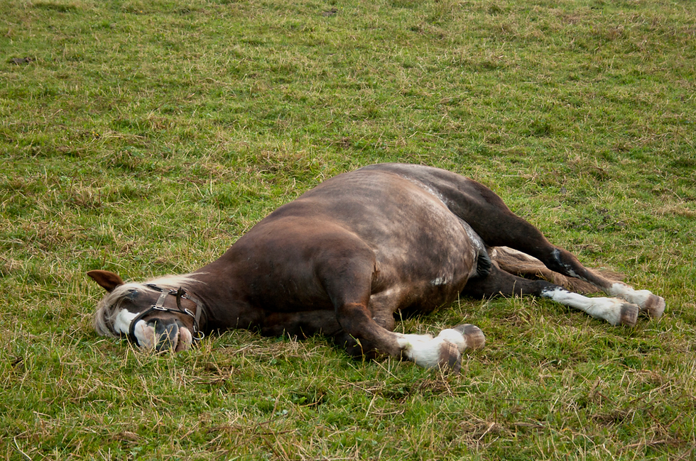
Colic is one of the most familiar and most feared health problems in horses. Many horse owners eventually encounter a horse that refuses a meal, paws at the ground, looks repeatedly toward its flank, lies down more than usual, or begins rolling. These behaviors are commonly described simply as “colic.”
Colic describes abdominal pain, not a single disease. The source of that pain may be as relatively minor as temporary gas accumulation or a change in intestinal movement, or as serious as a section of intestine that has twisted and lost its blood supply. Between those two extremes are impactions, intestinal displacements, inflammatory diseases, parasite-associated disorders, enteroliths, sand accumulation, gastric disease, intestinal tumors, and many other conditions.
This distinction is one of the most important things for a horse owner to understand.
Two horses may initially show almost identical signs of discomfort but have very different diseases developing inside the abdomen. Conversely, a horse with a life-threatening intestinal lesion does not always show dramatic pain.
Most episodes of colic do not require surgery, and many horses recover quickly with appropriate veterinary care. The challenge is recognizing the smaller proportion of horses whose disease is becoming dangerous. With some intestinal conditions, especially those that interfere with the blood supply to the intestine, hours can matter.
The purpose of this article is to provide a broad foundation for understanding equine colic: what the term means, why horses develop abdominal pain, how the major forms of colic differ, which horses require special consideration, what happens when surgery is needed, and what owners can reasonably do to reduce risk.
It is not intended to teach owners to diagnose individual intestinal lesions. That distinction belongs to the attending veterinarian. Instead, understanding the major disease processes allows an owner to recognize why apparently similar episodes of “colic” can have completely different outcomes.
If your horse is showing signs of colic now, contact your veterinarian immediately. Do not wait to identify the type of colic before requesting help. The sections below explain the diseases and decisions involved; they cannot establish a diagnosis for an individual horse.
Understanding Equine Colic
Why Colic Is a Syndrome Rather Than a Disease
A syndrome is a recognizable pattern of clinical signs that can have several underlying causes. A lesion is the specific abnormality responsible, such as an impacted colon or a strangulated segment of small intestine. Keeping those terms separate helps students distinguish what they observe from what has actually been diagnosed.
The veterinarian must determine whether the signs represent abdominal pain, whether the gastrointestinal tract is responsible, and what process is occurring. The immediate decision is often whether medical management is appropriate or whether the bowel needs surgical correction.
Sometimes the exact lesion is identified during examination. In other cases, the affected region and urgency become clear before the precise diagnosis, which may only be established at surgery. Treatment need not wait for a complete disease name when evidence already indicates compromised intestine.
Why the Horse Is Susceptible to Colic
The equine gastrointestinal tract is adapted to processing fibrous plant material through frequent feeding and hindgut fermentation. Natural grazing behavior combines movement with relatively small amounts of forage consumed over much of the day.
Managed horses may instead receive large meals, spend prolonged periods in stalls, eat substantial concentrate feeds, travel, and experience abrupt changes in water intake or routine. These differences can alter intestinal contents, movement, and microbial fermentation.
Anatomy contributes as well. The horse has a relatively small stomach and cannot effectively relieve substantial gastric pressure by vomiting. The small intestine includes long, mobile loops. The large colon is bulky and mobile, with changes in diameter and several bends. These features create opportunities for distention, impaction, displacement, and entrapment. Risk reflects the interaction of anatomy, disease, and management rather than a single flaw in the digestive system.
Digestive Anatomy That Explains the Different Types of Colic
Feed passes from the mouth through the esophagus to the stomach, then through the small intestine into the hindgut. The hindgut includes the cecum, large colon, small colon, and rectum.
The terms “small intestine” and “small colon” describe different structures. The small intestine lies between the stomach and cecum. The small colon lies much farther along the tract, after the large and transverse colons. Their diseases produce different examination findings and treatment needs.

The small intestine has three regions: the duodenum, jejunum, and ileum. The jejunum forms many mobile loops supported by mesentery, a sheet of tissue containing blood vessels, nerves, and lymphatic vessels. The ileum joins the cecum at the ileocecal junction. This narrow transition helps explain why disease around the ileum and tapeworm attachment sites receives particular attention.
The cecum and large colon support fermentation. Microorganisms break down fiber into products that the horse can absorb and use for energy. Normal digestion therefore depends on both the horse’s tissues and the microbial community within the bowel. Changes in diet can affect fermentation as well as the physical texture of intestinal contents.
The large colon follows a folded course through the abdomen. Its pelvic flexure makes a sharp turn and narrows, creating an important location for impaction. Portions of the colon have substantial freedom to move, which helps explain their susceptibility to displacement and twisting. Farther along, water absorption and the formation of fecal balls continue in the small colon. [1]
Did you know? A problem in the small intestine can threaten the stomach. When fluid cannot move forward, it may accumulate behind the obstruction and back up into the stomach, where pressure can become dangerous.
Anatomical directions also matter. “Proximal” generally means nearer the beginning of the intestinal tract, while “distal” means farther along it. “Dorsal” means toward the back and “ventral” toward the underside of the body. These terms describe locations, not the severity of a disease.
How Abdominal Pain Develops
Abdominal pain can develop through several mechanisms.
Distention is one of the most common. When gas or fluid stretches the stomach or intestine, tension develops in the intestinal wall and associated tissues. Even an otherwise healthy section of intestine can become painful when sufficiently distended.
Spasm or abnormal muscular contraction can also cause discomfort. This is one explanation for some episodes traditionally described as spasmodic colic.
An obstruction creates pain through a combination of distention, altered contractions, pressure within the bowel, and progressive disruption of intestinal function.
Inflammation causes another form of pain. The intestinal wall may become inflamed during enteritis or colitis, while inflammation within the abdominal cavity may cause peritonitis.
The most dangerous mechanism is intestinal ischemia.
Ischemia means inadequate blood supply to tissue. When a piece of intestine twists around its blood supply or becomes trapped through an anatomical opening, venous drainage may first become obstructed. The intestine becomes congested and swollen. Continued strangulation can then reduce or eliminate arterial blood flow.
Without adequate oxygen, intestinal cells begin to fail and die. The intestinal barrier becomes damaged, fluid moves into the bowel and abdominal cavity, inflammatory substances enter the circulation, and the horse can develop profound shock.
A strangulating intestinal lesion is therefore fundamentally different from an uncomplicated impaction. Both may obstruct the movement of intestinal contents, but the strangulating lesion also threatens the survival of the intestinal tissue itself.
The Difference Between Simple and Strangulating Obstruction
A simple obstruction blocks intestinal passage while initially leaving blood supply intact. An impaction or enterolith can act this way. A strangulating obstruction compromises both passage and circulation, placing intestinal survival at immediate risk.
“Simple” describes the mechanism, not a guarantee of safety. Persistent pressure can eventually injure the bowel wall. Blood supply can also fail without a twist, as in some parasite-associated vascular lesions. Students should therefore ask three separate questions: Is passage blocked? Is circulation impaired? Is the intestinal wall still viable?
Early referral can determine whether the surgeon can restore normal position or must remove dead bowel. As injury progresses, the consequences extend beyond the affected segment to fluid loss, systemic inflammation, and shock.
Recognizing Colic and Responding Early
Recognizing Colic
Colic often begins quietly. A horse may leave a meal unfinished, stand apart from the herd, repeatedly lie down, or look toward its abdomen before more obvious pain develops.
Pain behavior varies. Some older horses and donkeys show subtle discomfort despite serious disease.
Owners should therefore pay attention to changes from the individual horse’s normal behavior rather than waiting for spectacular signs.
Signs commonly associated with abdominal pain include:
Reduced appetite or complete refusal to eat.
Pawing, flank watching, or kicking or biting toward the abdomen.
Stretching repeatedly, lying down more than normal, or repeatedly getting up and down.
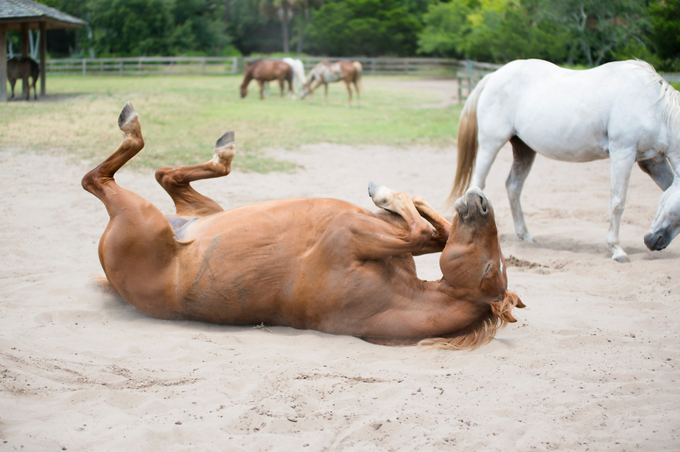
Rolling.
Sweating without an obvious environmental or exercise-related reason.
Restlessness or pacing.
Reduced manure production or unusually dry or loose manure.
Abdominal enlargement.
Depression or unusual quietness.
Rapid breathing.
Increasingly violent or uncontrollable behavior.
No single sign identifies the underlying disease.
Manure production must be interpreted alongside the other findings.
Likewise, the disappearance of pain is not always proof that the horse has recovered. In catastrophic gastrointestinal rupture or advanced shock, a severely painful horse may become quiet and depressed because the disease has progressed rather than improved.
The direction in which the horse is changing is therefore extremely important.
Did you know? Passing manure does not rule out an intestinal obstruction. Material already beyond the blockage may continue to leave the digestive tract. Recovery must be assessed from the whole horse, including comfort, appetite, circulation, and examination findings.
Learn to assess your horse before an emergency through our guide to The Horse’s Vital Signs. A record of the horse’s usual resting measurements makes later changes easier to describe.
When Colic Becomes an Emergency
Every suspected episode of colic deserves prompt communication with a veterinarian. The urgency becomes particularly high when pain is severe, keeps returning, or progresses despite initial veterinary treatment.
A horse that cannot be kept safely from violent rolling or thrashing requires immediate attention. So does one that becomes increasingly depressed, develops marked abdominal enlargement, has reduced manure production together with continuing discomfort, becomes progressively sweaty or weak, or shows other evidence of circulatory deterioration.
A horse that appears briefly comfortable and then becomes painful again also deserves particular attention. Recurrent pain can occur when an analgesic effect wears off while the underlying intestinal lesion remains.
The most important owner contribution during a colic episode is observation and communication. Tell the veterinarian when the horse was last known to be normal, along with:
When it last ate and drank.
Recent manure production.
Changes in feed or water.
Recent travel.
Exercise or housing changes.
Previous colic episodes.
Previous abdominal surgery.
Reproductive status.
Parasite-control history.
Any medication already given.
Accurate information from the owner can materially affect veterinary decision-making.
Owners should not delay veterinary assessment while attempting to determine which form of colic the horse has.
What to Do While Waiting for the Veterinarian
The first response should make veterinary assessment faster and keep both the horse and people safe. Call before trying home remedies, and follow the attending veterinarian’s directions for that particular horse. An apparently mild episode can change while help is on the way.
Remove feed and prevent grazing. Do not offer grain, hay, treats, bran mash, or other food to test whether the horse is improving. Ask the veterinarian how to manage water; do not force drinking or administer liquids by mouth.
Choose a safe area. If the horse can be moved safely, use an uncluttered enclosure with suitable footing. Avoid tight spaces where a painful horse could trap a handler.
Use walking cautiously. Brief, quiet walking may help some horses if they are comfortable enough and the veterinarian advises it. Stop if the horse becomes tired, more painful, weak, or unsafe to handle.
Allow quiet rest. A horse lying quietly does not need to be repeatedly forced to its feet. Violent rolling and thrashing create injury risks, but handlers should not endanger themselves trying to control an uncontrollable horse.
Record changes. Note the time, behavior, manure production, and any measurements you can obtain safely. A short video may help describe intermittent signs, provided recording does not delay the call.
Prepare for referral. Locate the trailer, driver, destination, and relevant records early if the veterinarian thinks transport may be needed. [2,3]
Never pour mineral oil into a horse’s mouth. Liquid can enter the airway and cause serious lung injury. Stomach tubing, rectal examination, abdominal puncture, and therapeutic rolling are veterinary procedures, not owner first aid. Do not attempt an enema in an adult horse or use human remedies to try to clear a suspected obstruction.
Give pain medication only on the veterinarian’s instructions, including the product, dose, route, and timing. Report anything already given, even if it was part of the horse’s usual treatment. Repeated medication may suppress pain while disease progresses, and excessive or inappropriate use can cause additional harm. Comfortable behavior after a drug is therefore only one observation, not proof of resolution.
Our Emergency Colic Kit and First-Hour Checklist can help organize emergency contacts, observations, and supplies before a crisis occurs.
Did you know? Rolling is usually a response to abdominal pain. The common claim that an ordinary roll causes the intestine to twist is not a sound reason to walk a sick horse to exhaustion. Safe handling and prompt veterinary attention are the priorities. [3]
Why Mild Signs Still Need Attention
Many episodes are mild and improve promptly with veterinary treatment, sometimes without a specific structural lesion being identified. However, “mild” describes the horse at that moment. It is not a final diagnosis.
Sustained improvement matters. A horse returning to normal comfort and function differs from one that needs repeated intervention to remain comfortable. Persistent or returning pain warrants reassessment even when the individual episodes appear modest.
Gas Colic and Impactions
Gas and Spasmodic Colic
Gas colic is one of the most common explanations for mild abdominal pain.
Gas is normally produced during gastrointestinal fermentation. The problem develops when excessive gas accumulates, gas is not moved normally through the intestine, or intestinal contractions become abnormal.
Dietary change, altered fermentation, changes in intestinal motility, confinement, changes in activity, or another developing intestinal problem can contribute.
The term spasmodic colic is generally used when abnormal intestinal contractions are thought to be contributing to pain. In practice, gas accumulation and altered motility may occur together.
Most uncomplicated cases resolve without surgery. However, substantial gas distention can also occur secondary to a mechanical obstruction or displaced colon. For this reason, “gas” should not automatically be interpreted as harmless.
Increasing abdominal enlargement or persistent pain warrants reassessment.
Impaction Colic
An impaction occurs when gastrointestinal contents become sufficiently firm, dry, bulky, or compacted that normal movement through the intestine is impaired.
Impactions can occur in several areas, but the large colon is especially important. Factors that can contribute include:
Reduced water intake.
Changes in forage.
Poor-quality or poorly digestible forage.
Prolonged stall confinement.
Sudden reduction in exercise.
Dental problems.
Other factors affecting ingestion, hydration, or gastrointestinal movement.
Winter is often associated with concern about impaction. Several things may change simultaneously: horses may drink less, eat more dry conserved forage, move less, and spend more time confined.
Large-colon impactions often develop gradually. Early discomfort may be mild and intermittent. Some horses remain reasonably bright between episodes of pain.
Many impactions respond to medical management, particularly when recognized before severe distention or secondary complications develop.

An impaction, however, should not be dismissed simply because the horse is not violently painful. A persistent impaction can enlarge, dehydrate further, alter intestinal motility, or contribute to displacement of the colon.
Large-colon impaction often involves the pelvic flexure, although other regions can be affected. Treatment commonly aims to restore hydration and soften intestinal contents while controlling pain. Resolution is judged by repeated examinations and sustained improvement. The passage of a little manure, or of a lubricant given during treatment, does not by itself prove that the impaction has cleared.
Ileal Impaction
The ileum is the final portion of the small intestine before intestinal contents enter the cecum. An ileal impaction obstructs this relatively narrow section with intestinal contents.
Historically, certain coarse forages and tapeworm infection have been associated with increased risk in some populations and geographical regions.
Early pain may be moderate. As intestinal contents and fluid accumulate in front of the obstruction, loops of small intestine become progressively distended.
An ileal impaction illustrates why the boundary between “medical colic” and “surgical colic” is not always immediate. Some cases resolve medically. Others do not, and persistent small-intestinal distention or deterioration can make surgery necessary.

Cecal Impaction
The cecum is a large fermentation organ located primarily on the right side of the abdomen.
Cecal impaction is particularly important because some affected horses show surprisingly little pain. The cecum can progressively fill and enlarge while clinical signs remain mild.
Recent confinement or reduced activity has been associated with cecal impaction, including in horses confined because of another medical or orthopedic problem.
The major concern is progression to cecal wall injury or rupture.
A horse that appears only mildly uncomfortable can therefore have a clinically important cecal lesion. This is one reason persistence of colic matters even when the apparent intensity of pain is low.
Veterinarians distinguish firm, dry cecal contents from cecal emptying failure, in which the organ may enlarge with comparatively fluid contents. These problems can look similar from outside but do not necessarily behave alike. Failure of the cecum to empty is especially concerning after hospitalization, anesthesia, or another illness. Continued mild signs in those settings deserve reassessment rather than repeated assumptions that reduced manure simply reflects eating less.

Small Colon Impaction and Fecaliths
The small colon is the portion of bowel where fecal balls are formed before manure enters the rectum.
Impaction here produces a distal obstruction and may result in reduced or absent manure production and progressive abdominal discomfort.
Small-colon impaction can occur in adult horses but is particularly noteworthy in miniature horses. Young miniature horses are also predisposed to fecaliths, which are firm masses of fecal material capable of creating complete obstruction.
Complete small-colon obstruction may require surgery, especially when continued distention threatens the bowel wall.

Sand and Intestinal Stones
Sand Enteropathy
Enteropathy means disease of the intestine. In sand-associated disease, the large colon is particularly important. Horses living in sandy environments may ingest sand while grazing or eating feed from the ground.
Small quantities can pass through the gastrointestinal tract, but repeated ingestion can allow substantial amounts to accumulate, particularly within the large colon.
Sand can cause several problems. Accumulated sand can irritate the intestinal lining, interfere with normal movement, contribute to impaction, and produce recurrent or acute colic. Some affected horses develop diarrhea or weight loss as well.
Risk depends on management as much as geography. Feeding directly on bare sandy soil increases exposure. Maintaining adequate forage and reducing opportunities for soil ingestion are important preventive principles.
Psyllium can be part of veterinary treatment, sometimes combined with other measures. Results depend on the amount of sand, the treatment protocol, and whether ingestion continues. A routine supplement should not be assumed to remove a clinically important accumulation. Prevention begins with reducing exposure to sandy soil. [4]
Abdominal radiographs can help identify and assess sand accumulation. A manure sedimentation test may show that sand is passing, but a negative sample does not reliably exclude retained sand, and a positive sample does not measure the total burden. Clinical findings and imaging are more useful than deciding from one manure sample alone. Severe obstruction or associated displacement may require surgery.
Did you know? Sand in the intestine does not always produce immediate colic. Recurrent discomfort, loose manure, or weight loss may precede an acute episode. This makes feeding practices and the horse’s longer history relevant even when the current episode seems sudden.
Enterolithiasis
An enterolith is a mineral concretion, or intestinal stone, that usually forms in the large colon. Minerals accumulate around a small central object, such as a pebble or other indigestible material. Equine enteroliths commonly contain struvite, a magnesium ammonium phosphate mineral.
Layers of minerals accumulate around a central object and gradually form a stone-like mass. Enteroliths can remain present for a long time without causing obvious illness.
Problems develop when an enterolith partially or completely blocks a narrower section of bowel. Horses may develop intermittent episodes of colic as the enterolith temporarily obstructs and then moves. Eventually a complete obstruction can occur.

Enterolithiasis is strongly geographical. It has historically been reported particularly frequently in California and certain other western regions of the United States. Diet and mineral environment appear to influence formation, and associations with diets containing substantial alfalfa have been reported, although development is likely multifactorial.
A horse living in an enterolith-prone region with repeated unexplained colic therefore presents a different risk profile from an otherwise similar horse in a region where enteroliths are uncommon.
Geography, Diagnosis, and Treatment of Enteroliths
California is a well-recognized region of increased enterolith occurrence, and cases also occur elsewhere in the United States and internationally. Regional case reports do not provide a dependable worldwide ranking of risk. Previous residence, forage history, and local veterinary experience are more useful than assuming the disease is confined to a short list of places.
Radiographs can identify some enteroliths, but detection depends on their location, the horse’s size, intestinal contents, and imaging equipment. A negative image does not exclude every stone. An enterolith causing obstruction generally requires surgical removal. Dietary recommendations after surgery should address that horse’s risk of recurrence without creating abrupt or inappropriate changes in the overall ration. [5]
Large Colon Displacements and Volvulus
Large Colon Displacements
The large colon occupies a substantial portion of the horse’s abdomen and has considerable mobility. Sometimes the colon moves from its normal position without twisting tightly enough to cut off its blood supply. This is called a displacement.
Two important examples are nephrosplenic entrapment, also called left dorsal displacement, and right dorsal displacement.
Nephrosplenic Entrapment
In nephrosplenic entrapment, a portion of the large colon moves upward on the left side of the abdomen and becomes trapped over the ligament between the spleen and left kidney.
Pain can be intermittent. Gas distention may occur, and some horses move through periods of discomfort and apparent improvement. Some cases can be corrected medically while others require surgery.
Recurrence can occur in some horses, which is why repeated episodes of the same displacement may lead to discussion of preventive surgical options.
Selected horses may be treated with veterinary medication, controlled exercise, or rolling under anesthesia. Suitability depends on the examination and the horse’s condition. These approaches should never be copied as home treatments, and unsuccessful medical correction should not be allowed to postpone necessary surgery.
Right Dorsal Displacement
In right dorsal displacement, the large colon moves abnormally around the right side of the abdomen.
The displacement can interfere with normal passage of intestinal contents and produce substantial gas accumulation or obstruction.
Like nephrosplenic entrapment, some right dorsal displacements resolve without surgery while others progress or fail to respond to medical management.
The distinction between a displacement and a volvulus is critically important because a displacement may leave intestinal blood flow relatively intact, whereas a strangulating volvulus can rapidly destroy the colon. A displacement can still become serious through obstruction, progressive distention, or pressure on tissues.
Large Colon Volvulus
Large-colon volvulus is among the most serious causes of equine colic.
A volvulus occurs when the large colon twists around its axis. If the twist is severe enough, it compresses the blood vessels supplying and draining the bowel. The colon rapidly becomes congested, swollen, and ischemic. Fluid and toxins move into the intestine and circulation, shock develops, and irreversible damage can occur within hours.
Affected horses frequently show acute, severe pain that becomes progressively difficult to control. Abdominal distention and cardiovascular deterioration may follow.
Large-colon volvulus is a true surgical emergency.
The chance of preserving viable colon is greatest when the horse reaches a surgical facility before extensive tissue death and systemic shock have developed. Delays can convert a correctable twist into irreversible intestinal injury.
Large-colon volvulus is particularly important in broodmares during the period around foaling, although it occurs in other horses as well.
Strangulating Obstructions and Internal Entrapments
Strangulating Small Intestinal Obstruction
A strangulating obstruction is one of the most time-critical forms of colic.
A segment of intestine becomes trapped or twisted in a way that obstructs both the intestinal lumen and its blood supply. Fluid and gas accumulate ahead of the obstruction, the affected bowel becomes swollen, and ischemic injury progresses.
Several different diseases can produce this same basic emergency.
Did you know? “Benign” describes how a tumor behaves as a growth, not every consequence it can cause. A benign fatty lipoma can still produce a fatal emergency if its stalk strangles the intestine.
Pedunculated Lipoma
A lipoma is a benign fatty mass. In older horses, some lipomas develop on long stalks within the abdomen. The mass itself is not usually harmful. The danger comes from the stalk. A mobile pedunculated lipoma can wrap around a loop of small intestine like a cord, strangling the bowel and cutting off its blood supply.
This is an important cause of surgical colic in geriatric horses.
An older horse with a strangulating lipoma may not display the dramatic pain expected from such a serious lesion. Subtle pain in an elderly horse therefore deserves particular respect.

Epiploic Foramen Entrapment
The epiploic foramen is a natural opening within the upper abdomen. A segment of small intestine can pass through this opening and become trapped. The entrapped intestine may then lose its blood supply.
Epiploic foramen entrapment is generally a surgical disease. Crib-biting or windsucking has repeatedly been associated with increased risk, although the behavior should be regarded as a risk association rather than proof of cause in an individual horse.
Small Intestinal Volvulus
A small-intestinal volvulus occurs when a section of intestine twists around its supporting mesentery.
The mesentery carries the blood vessels supplying the intestine, so twisting can produce rapid strangulation and ischemia.
Pain is commonly acute and severe, and emergency surgery is usually necessary.
Intussusception
An intussusception occurs when one section of intestine telescopes into an adjacent section. The involved segment can become obstructed and its blood supply compromised.
Jejunal intussusception affects the small intestine. Ileocecal intussusception involves the terminal small intestine and cecal region and is especially important in younger horses.
Some intussusceptions cause acute severe disease. Others can produce intermittent colic before becoming complete.
Inguinal Hernia
In stallions, small intestine can occasionally enter the inguinal canal and become trapped.
When the bowel is strangulated within the inguinal region, blood flow is compromised and emergency surgery is usually required.
Colts can also develop inguinal or scrotal hernias, although congenital hernias in young foals may behave differently from acquired strangulating hernias in adults.
Mesenteric Defects and Other Internal Entrapments
The intestine can occasionally become trapped through abnormal openings or tears in supporting tissues within the abdomen.
Examples include mesenteric rents, defects involving the gastrosplenic region, congenital remnants such as a mesodiverticular band, and abnormalities associated with a Meckel’s diverticulum.
These are considerably less common than gas colic or large-colon impaction, but they are important because they can create life-threatening strangulating obstructions.
Cecocolic Intussusception
The cecum itself can telescope into the adjacent colon. This is known as cecocolic intussusception. It can cause obstruction, compromise blood flow, and may require surgery.
Tapeworm infection has been associated with disease affecting the ileocecal and cecal regions, providing another example of how parasitism can contribute to specific forms of colic rather than simply producing a generalized “worm colic.”
Inflammation and Abnormal Intestinal Movement
Proximal Enteritis
Proximal enteritis, also called duodenitis-proximal jejunitis, is an inflammatory disease affecting the first portion of the small intestine. Its precise cause remains incompletely understood. Infectious organisms, toxins, diet, and other factors have been investigated, but no single cause explains all cases.
Inflamed small intestine stops moving normally and secretes or retains large volumes of fluid. Fluid then backs up toward the stomach. Affected horses may show abdominal pain, depression, fever or a history of fever, dehydration, and systemic inflammation.
One of the major clinical challenges is that proximal enteritis can resemble a mechanical small-intestinal obstruction, including a strangulating lesion. The distinction matters greatly. Proximal enteritis is usually managed medically, whereas a strangulating obstruction requires rapid surgery.
Some horses with proximal enteritis become critically ill and require intensive hospital treatment even though they do not need surgical correction of an obstruction.
Repeated gastric decompression and intravenous fluid support are often central to treatment. Some affected horses become more comfortable after fluid is removed from the stomach, but that response is not a reliable stand-alone test distinguishing enteritis from obstruction. Fever, ultrasound findings, reflux, abdominal fluid, and the clinical course must be considered together.
Colitis
Colitis is inflammation of the large colon. Affected horses may initially show abdominal pain before diarrhea becomes obvious. They can subsequently develop severe fluid loss, endotoxemia, systemic inflammatory disease, circulatory shock, and laminitis.
The term endotoxemia refers to the systemic effects associated largely with bacterial endotoxin crossing a damaged intestinal barrier or entering the circulation.
The healthy intestinal lining normally separates the enormous bacterial population of the bowel from the bloodstream. Severe inflammation damages that barrier. As a result, a horse with colitis may become critically ill even though there is no physical twist or obstruction requiring surgical correction.
Colitis illustrates an important principle: medical colic is not synonymous with mild colic. Some of the sickest equine gastrointestinal patients require intensive medical care rather than surgery.
Fever, depression, and diarrhea increase concern for inflammatory or infectious gastrointestinal disease. Because some causes of diarrhea can spread between horses, biosecurity may become an important part of management.
Infectious possibilities include Salmonella infection, certain clostridial diseases, equine coronavirus, and Potomac horse fever, depending on the region and clinical circumstances.
Noninfectious causes also occur, and testing does not identify a cause in every case. Until the veterinarian advises otherwise, separate a horse with acute diarrhea from other horses and use dedicated buckets and equipment. Tell the receiving hospital about diarrhea or fever before arrival.
Right Dorsal Colitis
Right dorsal colitis is a specific inflammatory disorder affecting the right dorsal colon.
It is classically associated with toxicity from nonsteroidal anti-inflammatory drugs, particularly excessive exposure or treatment of susceptible horses.
The intestinal lining becomes inflamed and ulcerated. Protein can be lost through the damaged bowel wall, and horses may develop abdominal pain, reduced appetite, weight loss, diarrhea, ventral edema, or low blood protein concentrations.
This condition is an important reminder that commonly used medications are not harmless. Pain medication for a colic patient should be used under veterinary direction rather than repeatedly administered in an attempt to suppress recurring pain without determining its cause.
Peritonitis
Peritonitis is inflammation of the lining of the abdominal cavity.
It may develop following gastrointestinal rupture, intestinal injury, abdominal infection, leakage after surgery, reproductive-tract disease, or other causes.
Clinical signs vary but may include abdominal pain, fever, depression, reduced appetite, reduced intestinal motility, and systemic illness.
Septic peritonitis, in which bacterial contamination is involved, can be particularly serious.
Peritonitis may itself produce ileus, creating further gastrointestinal distention and pain.
Ileus and Loss of Intestinal Movement
Ileus means severe reduction or absence of normal propulsive intestinal movement without a physical object necessarily blocking the lumen.
It may develop because of inflammation, severe systemic disease, electrolyte disturbances, intestinal manipulation during surgery, ischemic injury, pain, or other factors.
Postoperative ileus is an important complication following colic surgery, especially after small-intestinal disease.
When the small intestine stops moving, fluid accumulates, bowel loops distend, and stomach contents may back up. The horse can therefore continue to experience serious gastrointestinal dysfunction even after the original mechanical lesion has been corrected.
Equine Grass Sickness
Equine grass sickness is a disorder affecting the autonomic nervous system, including nerves controlling gastrointestinal function.
It occurs predominantly in certain geographical areas, particularly parts of the United
Kingdom and northern Europe, although disease has been recognized elsewhere.
Damage to the nervous system causes severe gastrointestinal dysfunction. Depending on the form of disease, horses may develop ileus, abdominal distention, difficulty swallowing, excessive salivation, weight loss, sweating, and other autonomic abnormalities.
Grass sickness is uncommon or absent in many regions, so geography is an important part of assessing risk.
The cause remains incompletely established. Acute and subacute forms generally carry a grave prognosis; selected chronic cases may survive with intensive nursing and nutritional support. Cases also occur outside northern Europe, including parts of South America. Grass sickness should not be used as a general label for any horse that becomes colicky after grazing. [6]
Stomach Disease, Rupture, and Other Abdominal Causes
Gastric Impaction
Although far less common than large-colon impaction, the stomach can become impacted with feed material.
Poor mastication, meaning how well food is chewed, unusual ingested material, altered gastric emptying, dehydration of stomach contents, and certain feeding behaviors or feeds have been associated with cases.
Gastric impaction may produce chronic or acute colic and can become dangerous if severe distention leads to damage or rupture.
Some cases occur alongside another intestinal lesion rather than as the sole cause of disease.
Equine Gastric Ulcer Syndrome and Colic
Gastric ulceration is common in certain horse populations, particularly performance horses exposed to intensive exercise and management conditions associated with prolonged gastric acid exposure.
Gastric ulcers can cause poor appetite, reduced performance, changes in body condition, and recurrent abdominal discomfort.
However, ulcers require careful interpretation. Because gastric lesions are common even among horses presented for unrelated problems, finding ulcers does not automatically prove that every episode of colic was caused by them.
Gastric disease becomes particularly relevant in horses with recurring signs rather than a classic acute intestinal emergency. Severe ulceration only rarely progresses to perforation, but gastric perforation is life-threatening.
The syndrome includes disease of the squamous, nonglandular lining and disease of the glandular stomach. These tissues have different protective mechanisms, so findings and treatment plans are not interchangeable. Gastroscopy allows the veterinarian to inspect the stomach lining and interpret lesions alongside the horse’s history. Suspected ulcers should not become a reason to overlook an acute intestinal emergency.
Cecal Perforation and Gastrointestinal Rupture
Any full-thickness rupture of the stomach or intestine allows gastrointestinal contents to contaminate the abdominal cavity. The resulting peritonitis is catastrophic.
Gastric rupture is particularly important because horses cannot effectively vomit to relieve excessive stomach pressure. Severe distention can arise from disease within the stomach itself or, more commonly, from fluid backing up from an obstructed or nonfunctioning small intestine.
Rupture can produce an apparent paradox: a horse that has been violently painful may suddenly become quiet. This is not necessarily improvement.
Once a distended gastrointestinal organ ruptures, internal pressure falls and some of the stretching pain disappears. At the same time, contamination of the abdomen leads to severe shock.
Profound depression after a period of severe colic is therefore an emergency.
Neoplasia and Abdominal Masses
Neoplasia means abnormal tissue growth and includes benign and malignant tumors. The pedunculated lipoma discussed earlier is benign but can cause mechanical strangulation. Malignant tumors, including lymphoma and some intestinal carcinomas, can infiltrate the bowel wall, obstruct passage, impair movement, or cause inflammation and protein loss.
The presentation may include recurring colic, progressive weight loss, poor appetite, or unexplained deterioration over weeks or months. Abdominal tumors become an important consideration when repeated discomfort remains unexplained, particularly when accompanied by weight loss or abnormal examination findings.
When Colic Comes From Outside the Gastrointestinal Tract
Not every horse displaying “colic behavior” has primary intestinal disease. Diseases of the reproductive tract, urinary system, liver, kidneys, abdominal muscles, or other organs may produce signs interpreted as abdominal pain. Pregnant and postpartum mares are especially important examples.
Uterine torsion can cause colic late in pregnancy. Hemorrhage from reproductive blood vessels can produce pain and shock. Postpartum mares can develop uterine tears, metritis, internal hemorrhage, or other complications.
Testicular disease can produce colic-like signs in stallions. An inguinal hernia containing trapped intestine, although located in the groin, remains an intestinal cause of colic.
Severe laminitis, neurologic disease, musculoskeletal trauma, and systemic illness can occasionally cause behavior that owners initially describe as colic.
For this reason, “the horse is colicky” should be viewed as an observation requiring investigation rather than a final diagnosis.
Veterinary Assessment and Treatment
What Happens During a Veterinary Colic Examination
The veterinarian selects tests to guide immediate decisions and may relieve pain or decompress the stomach before completing the examination. Not every horse needs every test, and diagnostic work should not delay necessary referral.
History and Physical Examination
The history establishes when signs began, how they have changed, and what conditions might influence the differential diagnosis. “Differential diagnosis” means the list of plausible causes being considered. A recently foaled mare, an older gelding, and a weanling may show similar pain but require different priorities.
The physical examination includes pain behavior, heart and respiratory rates, temperature, hydration, gum appearance, and intestinal sounds. Adult resting heart rate is commonly around 28 to 44 beats per minute, but individual baseline and context matter. A rising rate can accompany pain, dehydration, or circulatory compromise. A normal reading early in an episode does not rule out important disease. [7]
Gum color, moisture, and capillary refill help assess circulation. Gut sounds help assess activity, but the abdomen is not a simple “noise equals normal” system. Some obstructed horses still have sounds, and quiet sounds do not name the lesion. Repeated observations provide more useful information than one reassuring finding.
Nasogastric Intubation and Gastric Reflux
A veterinarian may pass a tube through a nostril, down the esophagus, and into the stomach. This allows assessment and removal of abnormal gastric fluid accumulation, called reflux. Decompression can relieve pain and reduce the risk of gastric rupture. Reflux often directs attention toward small-intestinal obstruction or poor movement, although it must be interpreted with the rest of the examination.

Tubing also provides a route for selected treatments, but fluid should not simply be added to a stomach that is already failing to empty. Tube position and the suitability of enteral treatment must be established by the veterinarian. [8]
Rectal Palpation and Abdominal Ultrasound
Rectal palpation allows the veterinarian to feel accessible structures through the rectal wall. Abnormal size, position, tension, or firmness can help identify impaction, displacement, or distended small intestine. Only part of the abdomen is within reach, so apparently unremarkable palpation cannot exclude all disease. The procedure requires training and appropriate restraint because injury is possible.
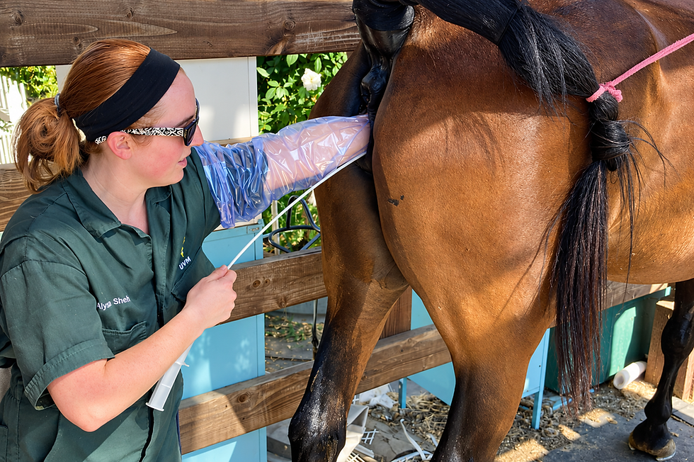
Ultrasound provides complementary information about intestinal position, wall thickness, fluid accumulation, and movement. It can reveal abnormalities that cannot be felt, but gas, depth, and access limit what is visible. The clinician interprets the pattern rather than expecting every lesion to appear as a single unmistakable image. [9]
Blood Tests and Abdominal Fluid
Blood testing helps evaluate hydration, inflammation, electrolyte balance, and organ function. Packed cell volume describes the proportion of blood occupied by red cells; interpretation alongside protein measurements is more informative than treating either value alone as a verdict on severity.
Abdominocentesis means collecting a sample of fluid from the abdominal cavity. Its appearance, cell content, protein, and other measurements can provide evidence of inflammation or bowel injury. Lactate in blood and abdominal fluid can support assessment of perfusion and intestinal compromise. Interpretation depends on timing and the overall case; an initially reassuring sample does not guarantee healthy bowel. [9]
Additional Tests and Repeated Assessment
Radiographs are especially useful for selected questions, including sand and enteroliths, rather than as a universal image of the adult horse’s entire intestine. Gastroscopy is useful when stomach disease is suspected. Recurrent cases may need further tests that are inappropriate or unnecessary during a rapidly evolving emergency.
Results guide the next decision: continue medical care, perform further assessment, or proceed to surgery. Repeated examinations reveal changes that one workup may miss.
How Medical Treatment Supports Recovery
Medical treatment is chosen for the disease and the horse’s condition. It may occur at the farm or require hospital care. The word “medical” describes the approach, not the seriousness of the illness; severe enteritis and colitis can require intensive treatment without a surgically correctable blockage.
Pain relief improves welfare and permits examination and safer handling. The veterinarian evaluates both the initial response and how long relief lasts. Recurring pain after appropriate medication is information that can change the treatment plan, not simply a reason to repeat the same dose indefinitely.
Fluid therapy supports circulation and corrects deficits. Depending on the lesion, fluids may be given intravenously or through a stomach tube. Enteral fluids can help selected impactions, but reflux or obstruction can make that route inappropriate. The plan must account for ongoing losses as well as the amount the horse has already lost.
Decompression and selected intestinal treatments address accumulation of gas, fluid, or dry contents. A lubricant does not untwist bowel or restore the blood supply to a strangulated segment. Similarly, medication that encourages motility is suitable only in selected circumstances after a veterinarian has considered obstruction.
Treatment of the underlying disorder may include measures for inflammatory disease, infection, parasites, or other identified causes. Antibiotics are not a routine solution for every colic. Their use depends on a specific indication, such as bacterial disease, sepsis, or perioperative needs. [10]
Monitoring continues while treatment is working. The team follows comfort, circulation, distention, reflux when relevant, hydration, and intestinal function. Failure to improve changes the plan. Medical management should have reassessment points, with clear instructions about which changes require an immediate call or referral.
After a horse improves, the veterinarian determines when and how feeding resumes. A hungry horse may look ready for its usual ration before the intestine is ready to manage it. Small, planned feedings and follow-up observations are safer than immediately replacing a missed day’s food. A horse that colicked at home still needs a recovery plan, even if it never required hospitalization.
How Veterinarians Decide Between Medical and Surgical Colic
Veterinarians combine pain pattern, circulation, intestinal distention and movement, reflux, laboratory findings, and response to treatment. Severe or returning pain despite appropriate analgesia raises concern, but no single measurement reliably determines the decision in every horse.
Repeated examinations reveal the direction of change. Improving comfort and intestinal function support a different plan from rising heart rate, worsening distention, and repeated pain. A horse can also require urgent surgery before every classic warning sign is present.
Referral provides continuous monitoring, intensive treatment, and access to surgery. Many referred horses recover with medical care, so referral itself should not be interpreted as a commitment to an operation.
Why Early Referral Matters
Earlier recognition and treatment can improve the opportunity for successful intervention. This is especially important for strangulating lesions.
An intestine deprived of blood cannot remain viable indefinitely. The longer strangulation persists, the greater the amount of tissue injury and the greater the systemic consequences.
Delayed treatment also increases the risk of postoperative ileus, adhesions, shock, and other complications.
Owners can improve preparedness before an emergency occurs by knowing where the nearest equine surgical facility is, having reliable transportation available, understanding insurance requirements if applicable, and deciding in advance whether surgery would be considered for each horse.
These decisions are much harder to make for the first time while a horse is actively deteriorating.
The practical discussion should include more than a yes-or-no decision about surgery. Ask what the examination suggests, what deterioration would look like, how transport will be managed, and what the likely options and costs are. Existing illness, welfare, expected recovery, and the owner’s ability to provide aftercare all matter. If treatment cannot relieve suffering or the lesion is not repairable, humane euthanasia may be the appropriate decision. Discussing these possibilities beforehand supports timely decisions without making assumptions about any owner’s resources.
Colic Surgery and Recovery
What Colic Surgery Is Intended to Accomplish
The broad purpose of colic surgery is simple even though the procedures themselves may be complex.
The surgeon enters the abdominal cavity to:
Identify the lesion.
Restore normal intestinal position and passage whenever possible.
Determine whether affected intestine is still viable.
Remove irreversibly damaged bowel when necessary.
Reduce contamination and distention.
The exact operation depends entirely on the disease.
A displaced colon may simply need repositioning. An enterolith may need removal. A strangulated segment of small intestine may require resection followed by reconnection of healthy intestine. A volvulus may require rapid untwisting and assessment of the colon.

The word “colic surgery” therefore does not describe one standardized operation.
Prognosis depends strongly on what disease is found, how long it has been present, how much intestine is damaged, the horse’s cardiovascular condition, and whether complications develop.
Understanding Surgical Survival Rates
Historically, owners often regarded colic surgery as a desperate last measure. Outcomes have improved substantially.
A 2023 multicenter study involving 451 surgical colic patients reported survival to discharge in 68.5% of all horses that underwent surgery in the study and approximately 80% among horses that survived anesthesia. The study involved three Italian referral centers and cases treated from 2018 through 2021. The different percentages describe different starting populations, not competing estimates for the same horse. [11]
These figures should not be applied mechanically to an individual horse. A horse undergoing surgery early for a correctable obstruction may have a very different prognosis from a horse arriving in advanced shock with extensive devitalized intestine.
The broader lesson is that surgery should not automatically be equated with hopelessness.
Likewise, advanced age alone is not a sound reason to assume that surgery cannot be successful.
Postoperative Recovery
Correcting the intestinal lesion is only the beginning of recovery. After abdominal surgery, veterinary teams monitor gastrointestinal function, circulation, hydration, pain, appetite, fecal production, the abdominal incision, and signs of infection or systemic inflammation.
Feeding is gradually reintroduced according to the type of intestinal disease, surgery performed, and return of gastrointestinal function. Horses that have experienced substantial intestinal damage may require a longer period before normal feeding can resume.
Owners should understand that complications do not necessarily mean that surgery has “failed.” Equine intestinal surgery involves tissue that may have been severely distended, inflamed, ischemic, or contaminated before the horse ever reached the operating room.
Postoperative Ileus
Postoperative ileus, often abbreviated POI, is one of the best-known complications after colic surgery. The intestine temporarily fails to regain effective movement. Fluid accumulates in the small bowel and stomach, causing distention and potentially recurrent pain.
Small-intestinal surgery, strangulating lesions, intestinal inflammation, and ischemia-reperfusion injury increase concern.
Some horses recover as intestinal function returns. Severe or persistent ileus can prolong hospitalization and increase the risk of additional complications.
Ischemia-Reperfusion Injury
Ischemia-reperfusion injury is additional tissue damage that can develop when circulation returns after inadequate blood supply. Restoring circulation remains essential, but previously injured cells and blood vessels may respond abnormally as oxygen returns.
Cells damaged during oxygen deprivation can generate inflammatory responses when oxygenated blood returns. Neutrophils, reactive oxygen species, inflammatory mediators, and vascular changes can contribute to additional tissue damage.
This helps explain why simply “untwisting the bowel” does not immediately return every severely affected horse to normal. The consequences of the original strangulation continue after circulation is restored.
Endotoxemia and Systemic Inflammation
Gram-negative bacteria normally live in the equine intestine. Components of their outer membranes include lipopolysaccharide, commonly called endotoxin. “Gram-negative” refers to a bacterial cell-envelope structure and staining classification. For colic, the relevant point is the presence of endotoxin in the outer membrane, not a rule that all such bacteria respond alike to antibiotics.
A healthy intestinal barrier limits systemic exposure.
Severe colitis, ischemia, intestinal damage, or other disease can compromise that barrier. Endotoxin and other inflammatory signals can then trigger a systemic response.
Affected horses may develop abnormal circulation, changes in gum color, fever, depression, organ dysfunction, and shock.
Endotoxemia is not a separate form of colic so much as a potentially serious consequence of gastrointestinal disease.
Adhesions
Healing tissues within the abdomen can occasionally attach to structures that would normally move freely past one another. These abnormal connections are called adhesions.
Small-intestinal disease and surgery carry particular concern because adhesions can tether or narrow sections of bowel and produce recurrent obstruction or colic. Not every adhesion causes clinical disease. The problem occurs when an adhesion alters intestinal position or movement enough to create pain or obstruction.
Recurrent colic after abdominal surgery therefore deserves evaluation rather than being assumed to be a routine consequence of having had surgery.
Incisional Complications and Hernias
The abdominal wall must heal after surgery just as the intestine does. Swelling around the incision can occur normally, but excessive pain, drainage, progressive swelling, separation, or other changes can indicate a surgical-site complication.
Infection can weaken the body wall and increase the later risk of an incisional hernia, in which abdominal contents bulge through a weakened area while remaining covered by skin.
Healing of the body wall is one reason postoperative exercise is restricted and gradually reintroduced.
Laminitis After Severe Gastrointestinal Disease
Laminitis is an uncommon but potentially devastating complication of severe gastrointestinal disease. Risk is greatest in horses experiencing severe systemic inflammation or endotoxemia, including some cases of colitis, enteritis, or peritonitis.
Veterinary teams therefore monitor the feet of seriously ill colic patients even when the original problem is entirely within the abdomen.
Long-Term Prognosis After Colic Surgery
Horses that recover sufficiently to leave the hospital often have a favorable long-term outlook. However, survival to discharge and survival years afterward answer different questions. Long-term studies also differ in follow-up duration, case selection, and how they count recurrent colic or other complications.
A 2026 study by Gandini and Giusto followed horses discharged after colic surgery. Of 203 discharged horses, follow-up information was obtained for 176, with a follow-up window of at least four years. Post-discharge complications were reported in 44.8%, and recurrent colic was the most common. The authors described long-term prognosis as favorable while emphasizing the importance of postoperative morbidity, meaning illness and complications.
The study was retrospective, relied on owner reports, and came from one center, which limits application to other populations. [12]
For an individual horse, the lesion, surviving bowel, cardiovascular condition, early recovery, and other health problems are more useful than an isolated overall percentage. A horse can survive and return to useful work while still requiring ongoing management. Conversely, survival alone does not describe comfort or quality of life.
Did you know? A reported surgical survival rate may include all horses taken to surgery, only horses that recovered from anesthesia, or only horses discharged from hospital. Always identify the starting group before comparing percentages.
Returning to Work
Many horses that recover from colic surgery return to their previous use.
Studies have documented successful return to pleasure riding, competition, breeding, and racing. The timeline depends on abdominal-wall healing, the surgery performed, postoperative complications, the horse’s body condition, and its intended athletic workload.
Colic surgery should therefore not automatically be viewed as the end of an athletic career.
Discharge instructions should specify feeding, medication, incision checks, exercise restrictions, and recheck appointments. Do not substitute a generic return-to-work calendar for the surgeon’s plan. Apparent energy and appetite can return before the abdominal wall has regained enough strength for unrestricted activity. Owners should report recurrent pain, wound drainage, progressive swelling, fever, or declining appetite promptly rather than waiting for the next scheduled visit.
Colic in Horses With Particular Risks
Colic in Foals
Foals are not simply small adult horses. Age changes the likely causes of abdominal pain, the useful diagnostic tests, and the consequences of missing a feed. Newborns have limited reserves and can deteriorate rapidly when they stop nursing or develop systemic disease.
In newborn foals, retained meconium can cause obstruction and straining. Meconium is the first fecal material, formed before birth from intestinal secretions and other material within the fetal gut. Passage normally begins soon after birth. Repeated straining, increasing distention, failure to nurse, or uncertainty about passage warrants veterinary assessment rather than waiting for a rigid deadline.
Other important causes include:
Enterocolitis: Inflammation involving small and large intestine, sometimes associated with infection or systemic illness.
Congenital abnormalities: Developmental problems that may block passage or prevent a segment from functioning normally.
Uroperitoneum: Urine accumulating in the abdomen because of a urinary-tract leak. This can cause distention and dangerous electrolyte disturbances while resembling intestinal disease.
Gastric and gastroduodenal disease: Ulceration or narrowing around the stomach and proximal intestine can affect feeding and emptying.
Intussusception or volvulus: Mechanical disease can obstruct passage and compromise the bowel’s blood supply.
Neonatal systemic illness: Sepsis and other serious diseases can produce abdominal signs and reduced intestinal movement.
A sick neonate may become unusually quiet, spend excessive time recumbent, lie on its back, grind its teeth, stop nursing, or repeatedly look toward its flank. These signs cannot distinguish retained meconium from a more dangerous condition at home. Enemas should be used only as directed by the veterinarian; repeated or inappropriate administration can injure the foal.

As foals grow, the pattern changes. Ascarid impaction becomes more relevant, along with hernias and other intestinal disease. Around weaning, equine proliferative enteropathy caused by Lawsonia intracellularis is another consideration. This infection thickens the small-intestinal lining and can cause poor growth, weight loss, diarrhea, colic, and low blood protein. It requires a different investigation and treatment plan from a mechanical obstruction.
Ascarid Impaction in Young Horses
Parascaris species are large roundworms primarily important in foals and young horses. A heavy intestinal burden can physically obstruct the small intestine.
One particularly important situation occurs when a heavily parasitized young horse receives an effective deworming treatment and large numbers of worms die over a short period. The resulting mass can contribute to obstruction.
This is one reason modern parasite programs emphasize surveillance and prevention of heavy parasite burdens rather than allowing large burdens to develop and then treating indiscriminately.
Anthelmintic, or dewormer, resistance has become a major international problem, particularly in equine nematodes, a group that includes gastrointestinal roundworms. Parasite control should therefore be based on current veterinary recommendations, fecal surveillance, age, farm history, and documented drug effectiveness rather than routine frequent rotation of dewormers without evidence.
Colic in Pregnant Mares
Pregnancy changes both the anatomy of the abdomen and the consequences of serious disease.
Pregnant mares can develop the same gastrointestinal disorders as other adult horses, including impactions, small-intestinal strangulations, large-colon displacements, and large-colon volvulus.
They also have pregnancy-specific causes of abdominal pain.
Uterine torsion is particularly important during later pregnancy. The uterus twists around its long axis, placing both mare and fetus at risk.
Hemorrhage into reproductive tissues and body-wall problems can also cause colic-like signs.
Serious maternal disease, endotoxemia, prolonged inability to eat, anesthesia, and surgery can threaten the pregnancy. Nevertheless, necessary treatment should not be delayed solely because the mare is pregnant. Failure to correct a severe intestinal lesion threatens both mare and fetus.
Colic in the Postpartum Mare
The period immediately after foaling has its own set of risks. Postpartum mares may develop:
Large-colon volvulus, in which the colon twists.
Colonic displacement, in which the large colon shifts from its normal position and becomes trapped or malpositioned.
Cecal disease, including impaction and perforation.
Small-intestinal lesions.
Reproductive-tract injury.
Internal hemorrhage.
Uterine tears.
Metritis, a serious uterine infection and inflammation after foaling.
Maternal behavior can make pain less obvious because some mares remain attentive to their foal despite substantial illness.
Repeated observation of the mare after foaling is therefore important. Reduced appetite, repeated lying down, sweating, abnormal abdominal contour, weakness, unusually pale gums, or signs of pain should not simply be attributed to the normal aftermath of parturition.
Colic in Stallions
Stallions have the same gastrointestinal diseases as other adult horses but have an additional important risk: inguinal herniation. A loop of small intestine can enter the inguinal canal and become strangulated. Exercise, breeding, and individual anatomy may contribute.
Acute abdominal pain in a stallion, particularly when accompanied by scrotal enlargement or asymmetry, requires rapid veterinary evaluation.
Colic in Older Horses
Older horses deserve particular attention because serious disease may produce deceptively mild signs.
Pedunculated lipomas, the stalked fatty tumors discussed earlier, become more common with age and can strangulate either small intestine or small colon.
Older horses are also susceptible to large-colon impaction, especially when dental disease interferes with proper chewing or management changes reduce forage or water intake.
Tumors become more relevant as horses age.
Chronological age alone does not determine outcome.
Studies comparing geriatric and mature horses have shown that appropriately selected older horses can survive colic surgery, including surgery for strangulating lesions. Age should be considered alongside the horse’s overall health, lesion, cardiovascular condition, and likely quality of life rather than used as the sole decision point.
Miniature Horses, Ponies, and Donkeys
Miniature horses have particular susceptibility to fecalith obstruction of the small colon, especially when young. A fecalith is a hard, rock-like mass of dried manure or ingested material that can block the lower intestine.
Ponies can develop many of the same lesions as full-sized horses, including impactions and strangulating lipomas as they age.
Ponies, miniature horses, and donkeys are also especially susceptible to serious fat-mobilization disorders when illness causes prolonged loss of appetite.
Hypertriglyceridemia and hyperlipemia can become life-threatening complications of the original gastrointestinal disease.
Donkeys create another challenge because their pain behavior may be extremely subtle. A donkey with serious colic may simply become quiet, stop eating, or appear depressed rather than rolling violently. Delayed recognition can worsen prognosis.
Owners should therefore treat appetite change in donkeys with particular caution.
Did you know? In a donkey, loss of interest in food may be the most obvious sign of serious abdominal disease. Waiting for violent rolling can delay recognition. Reduced intake also increases concern for hyperlipemia, so nutritional support must be planned alongside treatment of the original illness.
Recurrent Colic and Parasite-Associated Disease
Recurrent and Chronic Colic
Some horses experience one episode of colic and never have another. Others develop repeated episodes. Recurrent colic deserves investigation because it can reflect an underlying problem rather than simple bad luck.
Potential causes include repeated large-colon displacement, enteroliths, gastric disease, intestinal adhesions, chronic inflammatory or infiltrative bowel disease, sand accumulation, parasitism, intestinal tumors, poor dentition, feeding-management problems, stereotypic behavior such as crib-biting, and abnormalities of intestinal motility.
Previous colic itself is a recognized risk marker for future colic.
This does not necessarily mean that the previous episode damaged the intestine. Some horses may simply retain the same anatomical, management, dietary, behavioral, or physiological factors that contributed to the first episode.
A useful long-term record includes dates of episodes, severity, duration, changes in feed or turnout, veterinary findings, final diagnosis when known, hospitalization or surgery, and management changes made afterward.
Patterns can become apparent only when several episodes are viewed together.
Parasites and Colic
Parasites remain relevant to equine colic, but the relationship is more complicated than the old idea that any horse with worms simply develops “worm colic.”
Different parasites are associated with different disease processes.
Tapeworms have been associated with disease near the ileocecal region, including ileal impaction and intussusception.
Ascarids can create physical small-intestinal obstruction in young horses.
Cyathostomins can contribute to inflammatory large-intestinal disease.
Historically, Strongylus vulgaris was a major cause of vascular and intestinal disease.
Modern anthelmintic use has greatly reduced the prevalence of Strongylus vulgaris in many managed populations, although changes in parasite-control strategies require continued surveillance. Anthelmintics are medicines used to destroy parasitic worms.
At the same time, widespread resistance to anthelmintic drugs now limits the effectiveness of several drug classes. The modern goal is therefore not “deworm as often as possible.” It is to control clinically important parasitism while preserving drug effectiveness.
That generally means an evidence-based program developed with the veterinarian using fecal egg counts, age and risk group, treatment-history information, and periodic evaluation of whether the chosen products remain effective on the farm.
Larval cyathostominosis develops when large numbers of small-strongyle larvae emerge from the intestinal wall and provoke inflammation. Affected horses may develop diarrhea, weight loss, protein loss, and colic. Strongylus vulgaris can damage intestinal arteries and contribute to nonstrangulating infarction, meaning tissue death caused by interruption of circulation without a physical twist. These are different mechanisms from an ascarid mass blocking the bowel. [13]
Fecal egg counts estimate shedding by egg-producing parasites; they do not measure every parasite stage or exclude all parasite-associated disease. A low result cannot rule out encysted larvae, and standard testing has limitations for tapeworm detection. Use the veterinarian’s testing and treatment plan rather than interpreting one count as proof that parasites are irrelevant.
Our Horse Deworming Schedule Guide and Equine Deworming Schedule Planner can help organize that discussion and record planned care. Neither should be used to choose an emergency deworming treatment for a colicky horse.
Reducing the Risk of Colic
Feeding and Colic Risk
Nutrition is one of the most important areas of colic prevention, but it is also an area where simplistic claims are common.
No single diet can prevent every form of colic. The strongest general principle is consistency.
Horses evolved to consume forage over much of the day. Abrupt changes in forage, concentrate, turnout, or overall feed intake can alter the intestinal environment and microbial population.
Higher concentrate intake has been associated with increased colic risk in several studies. Large amounts of rapidly fermentable carbohydrate entering the hindgut can alter microbial fermentation and intestinal contents.
Poor-quality forage has also been associated with colic. Coarse, poorly digestible material may contribute to impaction in susceptible horses.
Specific regional associations also exist. Coastal Bermuda grass hay has historically been associated with ileal impaction in the southeastern United States, while alfalfa-rich diets have been associated epidemiologically with enterolithiasis in regions where enteroliths are common. These are associations with particular disease patterns, not proof that either forage is universally unsuitable.
These associations do not mean that every horse eating these forages will develop colic.
The appropriate question is how the whole diet, region, individual horse, water intake, dentition, exercise, and management interact.
Evaluate forage quantity, quality, and suitability for the individual horse before adding concentrates or supplements. Weigh feed rather than assuming every scoop or flake contains the same amount. Introduce changes progressively when circumstances allow, including changes between hay batches. Even a feed sold under the same name can differ in moisture, fiber, or energy content.
Our Horse Nutrition Basics resources provide a starting point for reviewing the whole ration. The Hay Net and Slow Feeder Selector can help assess feeding arrangements, with attention to safe use and whether the horse can actually obtain enough forage.
Did you know? Fiber is a carbohydrate. The nutritional concern is not “carbohydrates” as one category, but the type, amount, digestibility, and rate at which different feeds enter the digestive tract.
Management research identifies associations rather than guarantees about what will happen to a particular horse. The strongest interpretation comes from considering the diet alongside water, activity, housing, and recent changes. [14,15]
Water Is Central to Gastrointestinal Health
Water consumption can vary substantially with temperature, diet, workload, lactation, and individual behavior. Horses eating dry hay generally require more drinking water than horses consuming lush pasture.
Reduced water intake has been associated with colic, particularly impaction.
Cold weather deserves attention because horses may change their drinking behavior at the same time that they are eating more dry forage and exercising less.
Fresh, clean, palatable water should therefore be continuously available whenever possible.
Owners should become familiar with an individual horse’s normal drinking behavior, especially during weather changes, travel, illness, or confinement.
Check the water source itself. An automatic drinker may appear functional while delivering poorly, and a clean bucket at feeding time does not establish how much a horse drank overnight. Herd competition, unfamiliar water during travel, freezing, and contamination can all interfere with access or consumption.
Salt and electrolyte planning should match the horse’s diet and losses rather than being used as an emergency colic remedy. Our Equine Salt and Electrolyte Calculator supports routine planning; a horse that is painful, dehydrated, or not drinking needs veterinary assessment.
Pasture, Turnout, and Stall Confinement
Epidemiologic studies have repeatedly identified management change as an important part of colic risk.
Increased stall confinement and decreased pasture exposure have been associated with some forms of colic, particularly simple colonic obstruction and distention.
The relationship is unlikely to result from one factor alone.
A confined horse may move less, drink differently, receive a different diet, experience altered feeding intervals, and undergo behavioral stress simultaneously.
A horse suddenly confined for an orthopedic injury is a classic example. The horse that previously walked and grazed for many hours each day may abruptly begin standing in a stall and eating dry forage.
Appetite, drinking, and manure production deserve close attention during these transitions.
Exercise and Changes in Routine
Exercise itself is not simply good or bad for the gastrointestinal tract.
The more consistent finding is that changes in routine can matter. A horse moved abruptly from regular exercise to stall rest, or from light work to intense training, may experience simultaneous changes in feeding, water consumption, turnout, stress, and gastrointestinal motility. Intensive athletic management is also associated with gastric ulcer disease.
Management changes should therefore be made progressively whenever circumstances allow.
Dental Disease and Colic
Efficient chewing is the first stage of digestion. A horse with significant dental disease may swallow larger or less effectively processed feed particles. Poor dentition can also reduce total feed intake and alter the types of forage a horse is able to consume.
Longer intervals since dental care have been associated with some impaction-type colics.
Older horses deserve special attention because dental wear or tooth loss may make long-stem forage increasingly difficult to process. In these animals, appropriate alternative fiber sources may become an important part of maintaining gastrointestinal health.
The Senior Horse Hay Replacer Planner can help organize questions about alternative fiber sources when chewing becomes difficult. Selection, preparation, and amounts should be reviewed with the care team. Substituting an unsuitable feed or changing the entire ration suddenly can create new problems while trying to address the original one.
Crib-Biting and Windsucking
Crib-biting and windsucking have been associated epidemiologically with colic.
The association is particularly notable for epiploic foramen entrapment, although the biological explanation remains incompletely understood.
It is important not to confuse association with certainty of causation. A horse that cribs will not necessarily develop colic, and preventing the visible behavior does not necessarily eliminate the underlying physiological or management factors associated with it.
Nevertheless, stereotypic behavior should be recorded as part of the history of a horse experiencing recurrent abdominal disease.
Transportation
Recent travel has been associated with increased colic risk in some studies.
Several mechanisms may contribute simultaneously:
Decreased water intake.
Changes in feeding schedule.
Prolonged confinement.
Stress.
Reduced movement.
Exposure to unfamiliar environments.
Horses that repeatedly experience gastrointestinal problems around transportation warrant an individualized management plan developed with their veterinarian.
Geography Matters
Location changes the relative likelihood of particular diseases. Enteroliths are especially relevant in California, sand exposure depends on soil and feeding practices, and some forage-associated ileal impactions are reported more often in the southeastern United States. Grass sickness is particularly recognized in the United Kingdom and parts of northern Europe, with cases elsewhere. Parasite populations and drug resistance also vary. Tell the veterinarian where the horse previously lived, not just its current address.
A Practical Colic Prevention Program
Management can reduce some risks, but it cannot eliminate every cause of colic.
A carefully managed horse with excellent nutrition, water, parasite control, dentistry, and turnout can still develop a pedunculated lipoma, intestinal volvulus, congenital abnormality, tumor, or another unpredictable lesion.
Prevention should therefore be described as risk reduction rather than a guarantee.
A practical prevention program should:
Provide adequate forage.
Maintain reliable access to clean water.
Minimize abrupt changes in feed and routine.
Maintain appropriate turnout and activity.
Address dental disease.
Use evidence-based parasite control.
Reduce sand ingestion where relevant.
Monitor horses undergoing confinement or transportation.
Pay attention to risk factors particular to the individual horse and geographical region.
Owners should also keep records of previous colic. Horse Tracker and our Horse Information Sheet Guide can help organize the history for caregivers and the veterinarian.
A previous episode is itself one of the more consistent predictors of another episode.
Know Your Horse’s Normal
Many serious colic cases are first recognized because someone notices something small.
The horse does not finish breakfast.
There is less manure in the stall.
The normally social gelding stands quietly in a corner.
The old mare breathes a little faster and keeps looking at her flank.
The donkey stops eating.
None of these signs identifies a particular disease, but they can identify that something has changed.
Early recognition gives the veterinarian more options. This is particularly important with strangulating disease, where waiting for spectacular pain or profound shock can mean waiting until intestinal damage has progressed substantially.
Education and Continuing Care
Owner Education and Long-Term Follow-Through
Owners and daily caregivers usually see changes before the veterinary team does. The value of education is being able to notice, describe, and act on those changes without trying to name an intestinal lesion from behavior alone. Small changes in eating, manure, and behavior can provide the first warning.
Practice routine observations when the horse is well. Learn safe methods for checking vital signs, know where emergency contacts are kept, and make sure other caregivers can find them. Keep medication records clear enough that a substitute caregiver can tell what was given, when, and on whose instructions. Assign responsibility for checking intake when several people share care.
Our free Horse Ownership Foundations course provides a structured review of daily care and emergency preparation. Students seeking broader background can explore our Equine Study Kits. Understanding anatomy, behavior, and husbandry helps connect the observations made at the barn with the explanations given by the veterinarian.
After colic, review the discharge instructions with everyone responsible for the horse. Identify who monitors appetite, manure, water intake, and medication, and who calls if something changes. A well-organized record is particularly useful when the regular veterinarian and referral hospital both contribute to care.
Review risk factors without assuming that every episode represents an owner’s mistake. An anatomical entrapment, tumor, or sudden twist can occur despite careful management. Correct identified problems deliberately, and prioritize consistent care over unproven supplements.
Putting the Different Forms of Colic Together
Colic mechanisms often overlap. A strangulating lesion can cause distention, inflammation, ileus, and shock in the same horse. An initially uncomplicated impaction can become serious if it persists. A medically managed inflammatory disease may be more dangerous than a promptly corrected surgical obstruction.
The useful distinction is the process, its severity, and its response to treatment. No single behavior, vital sign, manure pile, or response to medication describes the whole case. Owners contribute by recognizing change and acting promptly; veterinarians determine the diagnosis and treatment. Repeated reassessment establishes whether the horse is recovering as expected or needs a different level of care.
Veterinary References
Merck Veterinary Manual. Overview of Colic in Horses.
UC Davis Center for Equine Health. Colic Happens.
UC Davis Center for Equine Health. 10 Things You Might Not Know About Equine Colic.
Merck Veterinary Manual. Sand Enteropathy in Horses.
UC Davis Center for Equine Health. Enterolithiasis.
Merck Veterinary Manual. Equine Dysautonomia. Updated September 2026.
American College of Veterinary Surgeons. Colic in Horses.
Merck Veterinary Manual. Colic in Horses: Horse Owners Version.
University of Minnesota. Large Animal Surgery Supplemental Notes. Field Diagnostics.
Reuss SM. Updates in the Medical Management of Colic: Moving Beyond Mineral Oil. AAEP Proceedings. 2014.
Spadari A, Gialletti R, Gandini M, et al. Short-Term Survival and Postoperative Complications Rates in Horses Undergoing Colic Surgery: A Multicentre Study. Animals. 2023;13(6):1107.
Gandini M, Giusto G. Evaluation of Long-Term Postoperative Morbidity and Survival After Equine Colic Surgery Using a Complication Severity Classification. Veterinary Record. 2026.
Nielsen MK. Diseases Resulting From Gastrointestinal Parasites in Horses. MSD Veterinary Manual. Reviewed October 2024.
Curtis L, Burford JH, Thomas JSM, et al. Risk Factors for Acute Abdominal Pain (Colic) in the Adult Horse: A Scoping Review of Risk Factors, and a Systematic Review of the Effect of Management-Related Changes. PLoS ONE. 2019;14:e0219307.
Gillen A, Archer DC. Epidemiology of Colic: Current Knowledge and Future Directions. Veterinary Clinics of North America: Equine Practice. 2023;39(2):157–174.
Frequently Asked Questions About Colic in Horses
1. What is colic in horses?
Colic means abdominal pain. It is a clinical sign or syndrome with many possible causes, including gas, impaction, inflammation, displacement, strangulation, parasites, sand, and intestinal stones. Urinary or reproductive disease can also produce abdominal pain. Identifying the cause requires veterinary assessment; the word “colic” alone does not establish severity or treatment.
2. What are the earliest signs of colic?
Early signs may include reduced appetite, leaving a meal unfinished, unusual quietness, pawing, flank watching, stretching, or repeated lying down. Some horses show severe pain quickly, while others remain deceptively quiet. Look for a change from the individual horse’s normal behavior and contact your veterinarian when colic is suspected.
3. When should I call the veterinarian for colic?
Call promptly for any suspected episode. Severe or returning pain, marked distention, weakness, abnormal gums, sweating, or worsening depression increase urgency. Foals, donkeys, recently foaled mares, and older horses may need particular vigilance. Do not wait for absent manure, violent rolling, or an abnormal heart rate before seeking advice.
4. Can a horse pass manure and still have colic?
Yes. Manure already beyond a developing obstruction can continue to pass, and many inflammatory or partial-obstruction conditions do not stop manure immediately. A manure pile is useful information but is not proof of recovery. The veterinarian considers sustained comfort, appetite, circulation, and examination findings as well as fecal production.
5. Should I walk a colicky horse or let it lie down?
Follow your veterinarian’s guidance. Brief walking may be reasonable when both horse and handler are safe, but do not force exercise or walk the horse to exhaustion. A horse resting quietly can generally remain lying down. Violent rolling creates injury risks; keep yourself safe rather than trying to physically overpower the horse.
6. Should I feed a horse that is showing colic signs?
Remove feed and prevent grazing while contacting the veterinarian. Ask for water instructions and never force fluids or oil into the horse’s mouth. Once the episode improves, the veterinarian should advise when feeding resumes, what to offer, and how much. Do not give a large meal to replace missed feed.
7. Does every horse with colic need surgery?
No. Most episodes are managed medically. Surgery becomes necessary for certain obstructions, strangulating lesions, and conditions that fail to respond appropriately to treatment. Referral also provides intensive monitoring and medical care, so going to a surgical hospital does not automatically mean an operation will be performed.
8. Can horses recover fully after colic surgery?
Many recover and return to their previous activities. Prognosis depends on the lesion, intestinal damage, circulation, other health problems, and postoperative course. Published survival percentages describe selected populations and cannot predict one horse’s outcome. Following individualized feeding, incision-care, and rehabilitation instructions is part of a successful recovery.
9. Can deworming prevent colic?
An effective parasite-control program reduces some parasite-associated risks, but it cannot prevent every form of colic. Unplanned treatment of a heavily parasitized young horse can contribute to obstruction. Use veterinary guidance, appropriate testing, and checks of drug effectiveness. Fecal egg counts do not rule out every parasite stage or associated disease.
10. Why does my horse keep getting colic?
Repeated episodes can reflect persistent management risks or underlying disease, including enteroliths, sand, gastric disease, displacement, adhesions, dental problems, inflammation, or tumors. Keep a record of episodes and treatment responses, and arrange a targeted investigation. Repeated pain medication without reassessment can delay recognition of a problem that needs different treatment.
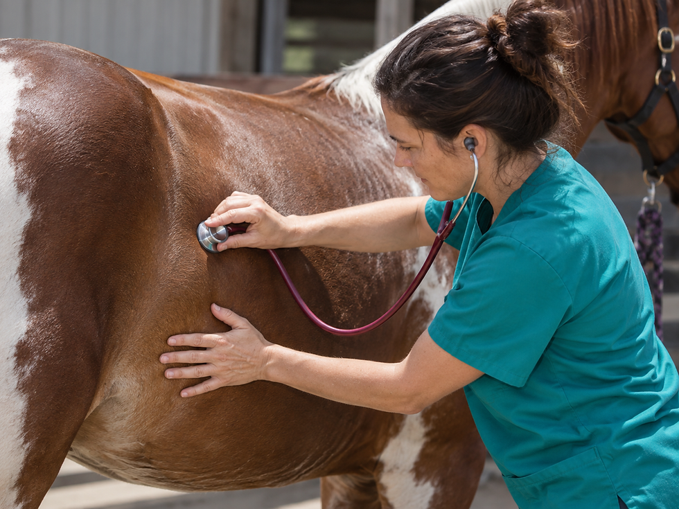


Comments