
Equine Body Muscle Score: A Professional Guide to Assessing Muscle Mass and Muscle Loss

Updated: Jul 28
Muscle Atrophy Scoring System, or MASS
Introduction
A horse’s muscle development is one of the most important indicators of strength, athletic readiness, soundness, recovery, and overall health. Muscle supports the skeleton, stabilizes joints, powers movement, protects soft tissues, and helps the horse perform ordinary activities such as rising, walking, turning, carrying a rider, and maintaining balance.
Equine Body Muscle Scoring is a practical system used to evaluate visible and palpable muscle over key regions of the horse. The goal is not simply to decide whether a horse “looks fit,” but to identify whether muscle is normal, reduced, uneven, or progressively wasting.
Don't confuse Equine Body Muscle Score with Body Condition Scoring. Body Condition Scoring (BCS) evaluates fat, while Body Muscle Scoring evaluates muscle. A horse can have an ideal body weight but still show muscle loss, especially along the neck, back, or hindquarters. Using both systems gives a clearer picture of the horse’s overall health, nutrition, strength, and physical condition. Read our article Body Condition Scoring: What It Is And How To Score Your Horse to learn more.
A research-based framework commonly used for this purpose is the Muscle Atrophy Scoring System, or MASS, which assigns a score from 1 to 4 to specific regions of the horse.
In this system 1 indicates no muscle atrophy, while 4 indicates severe muscle atrophy. The system was developed to provide a practical way to monitor skeletal muscle loss because more technical methods of measuring muscle mass can require specialized expertise, equipment, or expense.
Learn more about muscle anatomy with Horse Muscle Anatomy Explained: Major Muscle Groups, What They Do, And Why They Matter
Why Muscle Scoring Matters
Muscle loss can be gradual, subtle, and easy to overlook. A horse may continue eating normally and may even appear generally well while losing muscle over the topline, neck, or hindquarters. Regular muscle scoring helps identify these changes before they become severe.
A professional muscle score can help:
Establish a baseline for each horse
Monitor aging horses
Evaluate response to nutrition and exercise programs
Track rehabilitation after injury or illness
Identify muscle loss associated with pain, disuse, or disease
Detect asymmetry that may suggest lameness, neurologic disease, or uneven use
Improve communication between owners, veterinarians, farriers, trainers, and nutrition professionals
Loss of skeletal muscle mass may compromise both performance and welfare, which is why routine monitoring has practical value in horse management. In the MASS research, evaluations of the neck, back, and hindquarter regions showed acceptable reliability among trained raters, while the abdominal region was less reliable and is generally less useful as a primary scoring region.
The Overall Scoring System
The equine body muscle score is best understood as a regional scoring system, not simply one whole-horse number.
The most useful regions to assess are:
Neck
Back
Hindquarters
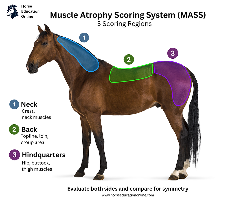
Each area is scored separately on a 1 to 4 scale:
This scoring approach allows the evaluator to describe where muscle loss is occurring, how severe it is, and whether it is symmetrical. A horse may score normally in the neck but show moderate loss through the back and hindquarters. Another horse may show severe loss on one side of the hindquarters only. These patterns are clinically important and should not be hidden by a single average score.
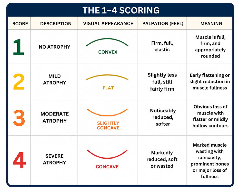
How Scores Are Achieved
Professional muscle scoring combines visual assessment and palpation. The evaluator looks at the horse, feels the muscle groups with a flat hand, compares both sides of the body, and assigns the score that best matches the observed shape, tone, and tissue depth.
1. Prepare the Horse
The horse should be standing square on level ground in good lighting. The coat should be clean enough to allow the evaluator to see surface contours. Thick winter hair, heavy blankets, poor lighting, uneven footing, or a tense posture can all make scoring less accurate.
The horse should be viewed from:
Both sides
The front
The rear
Slightly above or along the topline when appropriate
Photographs are helpful, but they should not replace palpation.
2. Evaluate Muscle Shape
A key principle of muscle scoring is the distinction between convex, flat, and concave muscle shape.
Convex
Convex muscle curves outward. This usually indicates normal or well-maintained muscle mass.
Flat
Flat muscle has lost some fullness. This may indicate mild or moderate muscle reduction, depending on the region and firmness.
Concave
Concave muscle curves inward. This is a stronger indicator of significant muscle loss, especially when paired with prominent bones or a rigid, soft, or poorly developed feel.
Current scoring guides advise evaluators to assess whether muscles are flat, concave, or convex, and to use a flat hand with light pressure when palpating the scored areas.
3. Palpate Muscle Tone
Muscle should not be judged by appearance alone. Palpation helps determine whether the tissue feels firm and elastic, soft, rigid, reduced, or uneven.
When palpating:
Keep the hand flat
Use light, consistent pressure
Feel both sides of the horse
Compare left and right muscle mass
Avoid digging with the fingertips
Note pain, tension, heat, or flinching
Firm, elastic muscle is generally a positive finding. Soft, rigid, or markedly reduced tissue may indicate poor muscle quality or muscle loss.
4. Score Each Region Separately
The neck, back, and hindquarters should each receive their own score. In professional recording, the score should be written by region, for example:
Neck: 1Back: 2Hindquarters: 3
This is more useful than writing only “overall score 2,” because it shows the distribution of muscle loss.
When left and right sides differ, record the asymmetry clearly:
Hindquarters: Left 4, Right 2
Marked asymmetry should be discussed with a veterinarian, particularly if it is new, progressive, or associated with weakness, lameness, poor coordination, or loss of performance.

Regional Scoring Guide
Neck
The neck is scored by looking at muscle fullness from the poll to the shoulder and by feeling the muscle along both sides of the neck.
The horse’s neck muscles can be thought of in three main groups:
Topline muscles, such as the splenius and semispinalis capitis, which lift and stabilize the neck.
Underside muscles, such as the sternomandibularis, longus colli, and longus capitis, which flex and lower the head and neck.
Side and shoulder-support muscles, such as the brachiocephalicus, trapezius, rhomboideus, and serratus ventralis cervicis, which help turn the neck, move the forelimb, and support the shoulder.
Study the Horse's Muscles with our Free Interactive Horse Muscles Tool
A healthy horse needs these muscles to work together so the neck can be strong, flexible, balanced, and comfortable.
Score 1: No Atrophy
The neck muscles are rounded and firm. The transition from neck to shoulder appears smooth. There is no obvious shelf, hollow, or loss of fullness.
Example: A mature riding horse in regular work has a smooth, rounded neck. The muscles feel firm and even on both sides. There is no depression where the neck meets the shoulder.
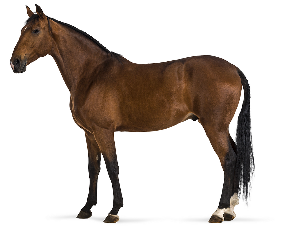
Score 2: Mild Atrophy
The neck begins to look flatter. A slight shelf may appear where the neck joins the shoulder. Muscle is still present but has lost some roundness.
Example: A horse returning from several weeks of reduced exercise has a slightly flatter neck than usual. The muscles still feel firm, but the transition into the shoulder is less smooth.
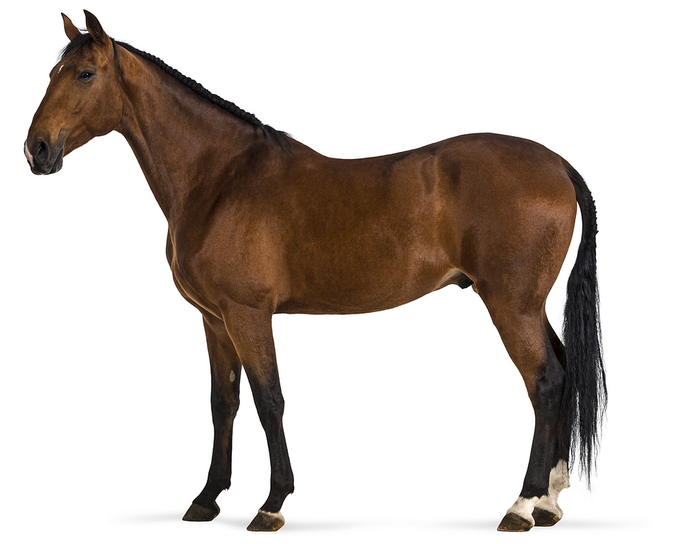
Score 3: Moderate Atrophy
The neck muscles are clearly reduced. They may appear flat or slightly concave. The neck may look narrower, and the shelf near the shoulder becomes more apparent.
Example: An older horse has a narrow neck with reduced muscle on both sides. The shoulder-neck junction is angular, and the muscles feel less elastic.
Score 4: Severe Atrophy
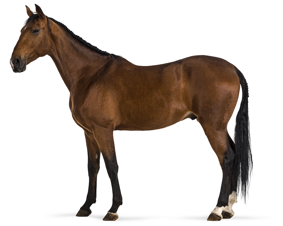
The neck appears hollow, thin, or sharply reduced. The muscle may feel rigid, very soft, or poorly developed. A prominent shelf may be visible where the neck meets the shoulder.
Example: A horse with progressive muscle wasting has deep concavity along the neck and a sharply defined shoulder junction. One side may be smaller than the other.

Back
The back is scored by assessing the muscles alongside the spine. These muscles are important for carrying posture, supporting the rider, transferring power from the hindquarters, and maintaining topline strength.
The main muscles of the horse’s back include the longissimus dorsi, illocostalis, spinalis, multifidus, latissimus dorsi , and the deep stabilizing muscles between the vertebrae. The abdominal muscles are also essential because they support the back from underneath.
A healthy horse’s back is not just strong. It must be flexible, stable, coordinated, and able to work with the neck, abdomen, pelvis, and limbs.
Score 1: No Atrophy
The back muscles are firm and full. The topline appears smooth. The muscles on either side of the spine are convex or flat but not hollow.
Example: A horse in steady work has a broad, firm back. The topline is smooth, and the muscles beside the spine are even.
Score 2: Mild Atrophy
The back muscles are slightly reduced. There may be mild flattening or early concavity. The top of the spine may become more visible, but the back still has reasonable muscle coverage.
Example: A horse after a short rest period shows mild loss of topline fullness. The muscles are slightly flatter than before but not deeply hollow.
Score 3: Moderate Atrophy
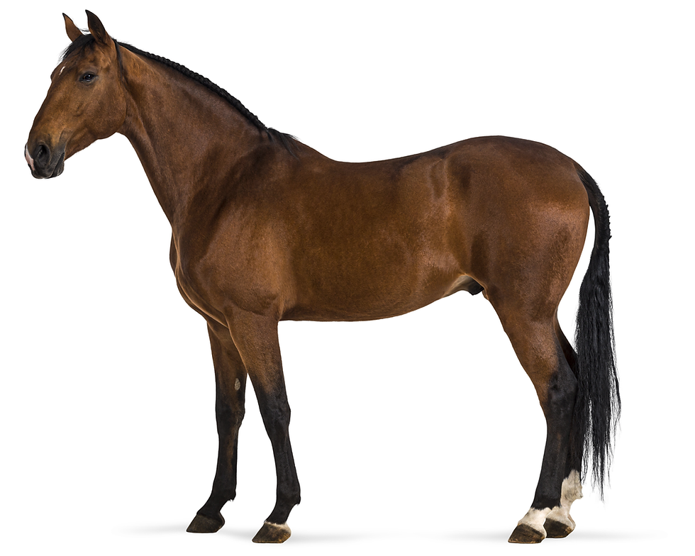
The back muscles are clearly reduced. Concavity may be visible alongside the spine. The bony structures of the topline become easier to see or feel.
Example: A horse that has been out of regular work for several months has a visible groove along the back, reduced muscle on both sides of the spine, and less ability to lift through the topline.
Score 4: Severe Atrophy
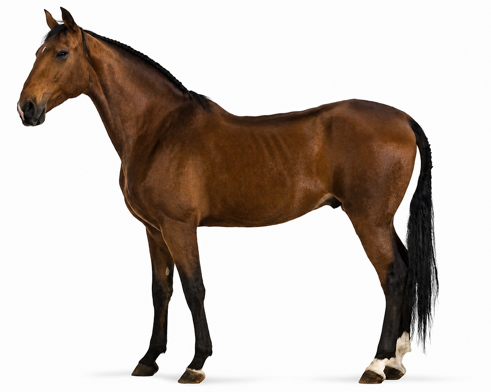
The back is deeply hollow. The spine is prominent, and the muscles on either side are markedly reduced. This score indicates significant muscle wasting.
Example: A chronically ill or severely deconditioned horse has a narrow, sunken back with very little muscle fullness along the spine.

Hindquarters
The hindquarters are among the most important areas to score because they contain large propulsive muscles. Loss here may affect power, balance, ability to rise, engagement, and athletic performance.
The major muscles of the horse’s hindquarters include the gluteal muscles, hamstrings, quadriceps, inner thigh muscles, deep hip stabilizers, iliopsoas, gastrocnemius, and digital flexor muscles.
A simple way to think about them is:
Gluteals and hamstrings create power.
Quadriceps stabilize and extend the stifle.
Iliopsoas and hip flexors bring the hind limb forward.
Adductors and deep hip muscles control limb position and stabilize the pelvis.
Gastrocnemius and flexor muscles help with hock action, push-off, and lower-limb support.
Together, these muscles allow the horse to move forward, carry weight, balance, turn, stop, jump, and perform athletic work.
Score 1: No Atrophy
The hindquarters are rounded, firm, and symmetrical. The croup and gluteal muscles are well filled. Bones of the pelvis are not unusually prominent.
Example: A fit horse has rounded hindquarters with even muscle development on both sides. The muscles feel firm and elastic.

Score 2: Mild Atrophy
The hindquarters begin to lose roundness. There may be slight flattening at the top or sides of the croup. Bony landmarks may be mildly easier to identify.
Example: A horse recovering from mild lameness has slightly flatter hindquarters, but the muscles are still present and gener
ally symmetrical.

Score 3: Moderate Atrophy
The hindquarters are visibly reduced. Muscles may be concave below the point of hip or over the croup. Pelvic bones become more noticeable.
Example: A horse with reduced work due to chronic soreness has flatter, narrower hindquarters. The croup lacks fullness, and the point of hip is more apparent.

Score 4: Severe Atrophy
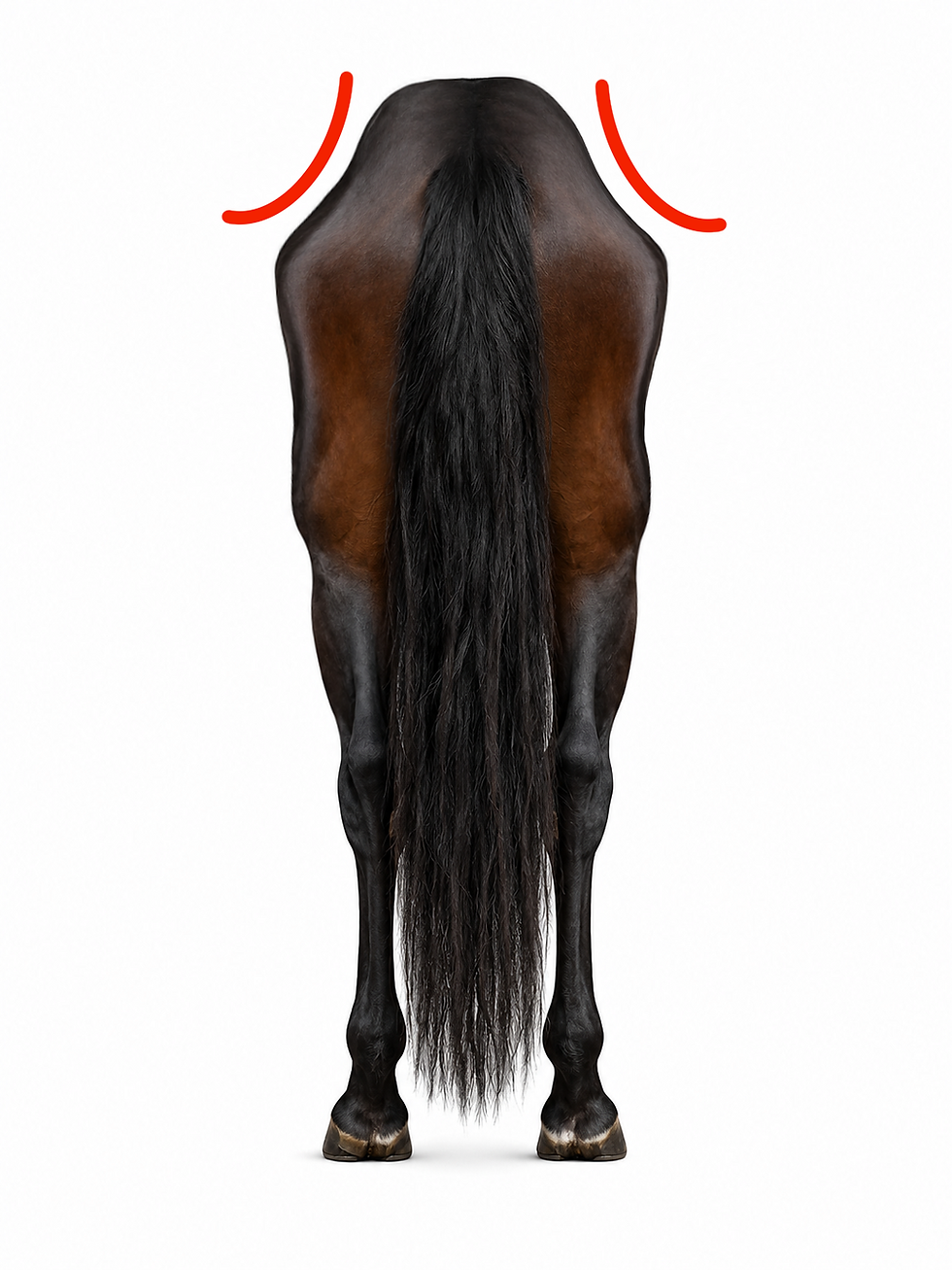
The hindquarters are deeply concave or markedly wasted. Pelvic bones are prominent. There may be obvious asymmetry between left and right sides.
Example: A horse with neurologic disease or severe disuse has one hindquarter much smaller than the other, with deep hollowing and loss of muscle mass.
How to Determine the Overall Result
Because muscle loss may be regional, the best professional approach is to report both regional scores and an overall interpretation.
Regional Score
Each area receives its own score:
Neck: 2
Back: 3
Hindquarters: 3
This tells the owner, veterinarian, or trainer where the problem is most evident.
Highest-Score Method
The highest regional score identifies the most severe area of muscle loss.
Example:
Neck: 1
Back: 2
Hindquarters: 4
Overall interpretation: Severe regional muscle loss, because one area scores 4.
This method is useful because it prevents serious localized muscle loss from being minimized.
Total-Score Method
For tracking progress over time, the three regional scores may also be added together:
Minimum total: 3
Maximum total: 12
Example:
Neck: 2 + Back: 2 + Hindquarters: 3 = Total 7
This can help monitor trends, especially when the same evaluator scores the same horse every 30 to 60 days. However, the total score should never replace the regional notes, because the pattern of muscle loss is often more important than the number alone.
Practical Scoring Examples
Example 1: Normal Muscle
Horse: 9-year-old gelding in regular work
Neck: Rounded and firm
Back: Smooth and well filled
Hindquarters: Symmetrical and muscular
Scores: Neck 1, Back 1, Hindquarters 1
Total: 3
Interpretation: No evident muscle atrophy.
Example 2: Mild Generalized Muscle Loss
Horse: 16-year-old mare returning from a period of reduced exercise
Neck: Slight flattening
Back: Mild topline reduction
Hindquarters: Slightly less rounded than previous photographs
Scores: Neck 2, Back 2, Hindquarters 2
Total: 6
Interpretation: Mild generalized muscle loss. This may reflect reduced workload, aging, early nutritional inadequacy, or a combination of factors.
Example 3: Moderate Topline and Hindquarter Loss
Horse: Mature riding horse after several months out of work
Neck: Normal
Back: Concavity along both sides of the spine
Hindquarters: Flattened croup and reduced gluteal fullness
Scores: Neck 1, Back 3, Hindquarters 3
Total: 7
Interpretation: Moderate regional muscle loss, most evident through the back and hindquarters. A progressive strengthening program and veterinary review for pain or lameness may be appropriate.
Example 4: Severe Asymmetrical Muscle Loss
Horse: Senior horse with declining coordination
Neck: Mild loss
Back: Moderate loss
Left Hindquarter: Severe wasting
Right Hindquarter: Mild to moderate loss
Scores: Neck 2, Back 3, Hindquarters Left 4 / Right 2
Interpretation: Severe asymmetrical muscle loss. This pattern warrants veterinary evaluation because uneven wasting may suggest neurologic disease, chronic lameness, or another significant medical issue.
What the Scores Mean Clinically
Score 1: No Atrophy
This is the desired finding. Muscle mass appears appropriate for the individual horse’s age, use, and management. Continue routine monitoring.
Score 2: Mild Atrophy
Mild atrophy is an early warning sign. It may reflect a recent decrease in exercise, early aging changes, poor protein intake, subtle pain, or recovery from illness. This score is often reversible when the cause is identified early.
Score 3: Moderate Atrophy
Moderate atrophy is clinically important. It suggests that muscle loss is no longer subtle. The horse may have reduced strength, reduced topline support, reduced hindquarter power, or impaired performance. A score of 3 should prompt a careful review of diet, workload, soundness, saddle fit, dental status, and overall health.
Score 4: Severe Atrophy
Severe atrophy indicates significant muscle wasting. It may be associated with chronic disease, pain, neurologic dysfunction, prolonged inactivity, malnutrition, or advanced age-related decline. A score of 4 should be treated as a serious finding, especially if the change is rapid or asymmetrical.
Rationale Behind the System
The rationale for equine body muscle scoring is based on three principles.
First, muscle loss affects function. A horse with reduced muscle may struggle to carry itself correctly, stabilize joints, generate power, or recover from work. Muscle loss may also affect welfare by making ordinary movement more difficult.
Second, muscle loss is often regional. A horse may lose muscle mainly through the back, one hindquarter, or one side of the neck. This pattern can provide valuable clues about use, pain, compensation, or disease.
Third, a practical scoring system allows repeated monitoring. Even when advanced imaging or laboratory testing is not available, a consistent visual and hands-on scoring method can identify trends over time. The MASS study found reliable scoring agreement for the neck, back, and hindquarter regions among evaluators, supporting the practical use of these areas for monitoring muscle atrophy in horses.
Best Practices for Accurate Scoring
For the most consistent results:
Score the horse in the same location each time.
Use good lighting.
Stand the horse square.
Evaluate both sides.
Palpate with a flat hand and light pressure.
Record each region separately.
Take photographs from consistent angles.
Recheck every 30 to 60 days when monitoring change.
Use the same evaluator whenever possible.
Record recent exercise, illness, lameness, diet changes, and medications.
If two evaluators disagree, they should review the horse together and discuss the specific region causing disagreement. Differences often occur when one person relies too heavily on visual appearance and the other places more emphasis on palpation.
Common Causes of Reduced Muscle Score
A declining muscle score may be associated with:
Reduced exercise or stall rest
Inadequate high-quality protein intake
Insufficient calories
Dental disease
Chronic pain
Lameness
Back pain
Poor saddle fit
Aging
Neurologic disease
Endocrine disease such as PPID
Chronic inflammatory or systemic illness
Recovery from injury or surgery
The score itself does not diagnose the cause. It identifies the presence, location, and severity of muscle loss so that the underlying reason can be investigated.
When to Seek Veterinary Evaluation
Veterinary evaluation is recommended when:
Muscle loss is rapid
A score increases from 1 or 2 to 3 or 4
Muscle loss is asymmetrical
The horse shows weakness or stumbling
The horse has difficulty rising
Performance declines unexpectedly
The horse appears painful during palpation
The topline or hindquarters deteriorate despite appropriate exercise
Muscle loss occurs with weight loss, dullness, fever, or abnormal behavior
A professional muscle score is most valuable when combined with a broader health assessment, soundness evaluation, nutrition review, and history of the horse’s workload.
Equine Body Muscle Scoring provides a structured, practical way to evaluate muscle mass and muscle loss in horses. By scoring the neck, back, and hindquarters separately on a 1 to 4 scale, owners and professionals can identify early changes, monitor trends, and recognize patterns that may point to disuse, pain, aging, nutritional problems, or disease.
The most important principle is consistency. The same method, the same regions, the same scoring scale, and careful written records allow subtle changes to become visible over time. A muscle score is not a diagnosis, but it is a valuable clinical and management tool that helps protect performance, comfort, and long-term health.
FAQ: Equine Body Muscle Scoring
What is Equine Body Muscle Scoring?
Equine Body Muscle Scoring is a visual and hands-on method used to evaluate muscle mass and muscle loss in specific areas of a horse’s body.
It helps identify whether muscle is normal, reduced, uneven or progressively wasting. Owners can use the Interactive Horse Muscles Tool to better understand the major muscle groups being assessed.
What is the difference between Body Muscle Scoring and Body Condition Scoring?
Body Muscle Scoring evaluates muscle, while Body Condition Scoring evaluates fat.
A horse may have an appropriate body weight and body condition while still losing muscle through the neck, back or hindquarters. Both systems should be used together for a more complete assessment. Read the guide to Body Condition Scoring in Horses for the fat-scoring method.
What is the Muscle Atrophy Scoring System?
The Muscle Atrophy Scoring System, or MASS, assigns each assessed region a score from 1 to 4.
A score of 1 indicates no muscle atrophy. A score of 2 indicates mild atrophy, 3 indicates moderate atrophy, and 4 indicates severe muscle atrophy.
Which areas of a horse should be scored for muscle loss?
The three main regions are the neck, back and hindquarters.
Each area should receive its own score because muscle loss may affect one region more severely than another. Recording only one overall number can hide important regional or one-sided muscle loss.
How do you assess a horse’s muscle score?
Stand the horse square on level ground in good lighting. View the horse from both sides, the front and the rear, then palpate the muscles using a flat hand and light, consistent pressure.
Evaluate whether the muscle shape appears convex, flat or concave, compare both sides and record each region separately. The interactive horse anatomy guide can help identify the relevant body regions.
What do convex, flat and concave muscles mean?
Convex muscle curves outward and usually indicates normal or well-maintained muscle mass.
Flat muscle has lost some fullness and may indicate mild or moderate reduction. Concave muscle curves inward and is a stronger sign of significant muscle loss, particularly when bones become prominent or the tissue feels poorly developed.
What causes muscle loss in horses?
Possible causes include reduced exercise, stall rest, inadequate protein or calories, dental disease, chronic pain, lameness, back pain, poor saddle fit, aging, neurologic disease, PPID, chronic illness and recovery from injury or surgery.
A muscle score does not diagnose the cause. It identifies where muscle loss is occurring and how severe it is so the underlying problem can be investigated. Owners should also review the basics of equine nutrition when evaluating diet-related muscle loss.
How often should you score your horse’s muscle mass?
When monitoring changes, the article recommends reassessing the horse every 30 to 60 days.
Use the same location, lighting, method and evaluator whenever possible. Consistent photographs and regional records make gradual changes easier to identify.
When does muscle loss require veterinary evaluation?
Contact a veterinarian when muscle loss is rapid, asymmetrical or progresses from mild to moderate or severe.
Veterinary assessment is also recommended when muscle loss occurs with weakness, stumbling, difficulty rising, pain, declining performance, weight loss, fever, dullness or abnormal behaviour. Records in Horse Tracker can help document regional scores, photographs, exercise changes, lameness, illness, diet and veterinary findings over time.


Comments