
The Equine Elbow; Anatomy, Function, and Pathology


The equine elbow connects the humerus of the upper forelimb with the radius and ulna of the forearm. This strong, stable joint allows the limb to bend during movement and support the horse’s weight when the hoof is on the ground. Its function depends on the coordinated work of bones, cartilage, ligaments, muscles, tendons, and nerves.
Elbow disease is less common than many causes of lower limb lameness, but injuries in this region can seriously affect a horse’s ability to stand, walk, and perform. Some problems produce obvious swelling or a characteristic dropped elbow. Others cause subtle changes in stride that require a thorough veterinary examination to identify.
Understanding the elbow’s anatomy helps owners and students distinguish a superficial swelling from a potentially serious joint, bone, muscle, or nerve injury.
Where Is the Horse’s Elbow?
The elbow sits high on the forelimb, beside the chest and below the shoulder. The prominent projection at its back is the olecranon, commonly called the point of the elbow.
The elbow should not be confused with the horse’s knee. The joint traditionally called the knee is the carpus, which sits farther down the forelimb and corresponds anatomically to the human wrist.
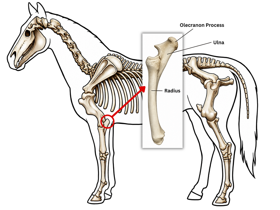
The region between the elbow and carpus is the antebrachium, or forearm. Familiarity with these landmarks makes it easier to describe an injury accurately and understand a veterinarian’s findings.
Use our Interactive Horse Anatomy Guide and Interactive Horse Skeleton to locate the elbow within the complete forelimb.
Did you know?A horse’s elbow corresponds to your elbow, while its front “knee” corresponds to your wrist. Much of the horse’s lower limb consists of structures equivalent to the bones of your hand and a single finger.
Anatomy of the Equine Elbow
The Three Bones
The elbow is formed by the humerus, radius, and ulna. Their shapes allow considerable bending and straightening while restricting sideways movement.
The humerus extends from the shoulder to the elbow. Its lower end has rounded articular surfaces, collectively forming the humeral condyle, that meet the forearm bones. The medial and lateral epicondyles provide attachment sites for ligaments and muscles. At the back, the olecranon fossa accommodates the anconeal process of the ulna during extension.
The radius is the main weight-bearing bone of the forearm. Its broad upper surface articulates with the humerus and transmits substantial loads through the limb.
The ulna is reduced compared with the human ulna and is extensively fused with the radius in the mature horse. Its upper portion forms the olecranon, the trochlear notch, and the anconeal process. The olecranon provides the attachment for the triceps, making it essential to the mechanism that extends the elbow.

For a broader skeletal overview, read Horse Skeleton Anatomy Explained. The Bones of the Horse Flashcards and Bones of the Thoracic Limb Poster provide additional visual references.
Articulations and Movement
The elbow contains two principal articulations:
Humeroradial articulation: Between the humerus and radius, forming the main weight-bearing interface.
Humeroulnar articulation: Between the humerus and ulna, contributing to joint stability and movement.

These articulations share a joint capsule and synovial cavity. The elbow functions primarily as a hinge joint, allowing flexion and extension. Flexion bends the joint; extension straightens it.
The close relationship between the radius and ulna greatly limits forearm rotation. Horses cannot turn the forearm in the way people rotate a hand from palm up to palm down.
Cartilage, Joint Capsule, and Synovial Fluid
Smooth articular cartilage covers the surfaces where the bones meet. It reduces friction and helps distribute pressure.
A fibrous joint capsule encloses the joint. Its inner synovial membrane contributes to the production and maintenance of synovial fluid, which lubricates the joint and supplies nutrients to the cartilage.
Healthy cartilage has no direct blood supply. Its maintenance depends partly on nutrient exchange with synovial fluid, supported by normal joint loading and movement.
Our article The Basics of Joints, With Notes on Arthritis explains these structures in greater detail. The Joint Anatomy Flashcards are useful for reviewing the terminology.
Collateral Ligaments
Strong collateral ligaments support both sides of the elbow:
The medial collateral ligament supports the inner side.
The lateral collateral ligament supports the outer side.

They connect the humerus with the forearm bones and limit abnormal sideways movement. Their stabilizing function allows the joint to carry weight while moving mainly in a forward-and-backward plane. CVM Large Animal Anatomy
Muscles That Control the Elbow
Triceps Brachii: The Main Extensor
The triceps brachii occupies much of the region behind the humerus. In horses, it has three heads: long, lateral, and medial. An accessory head, present in some other domestic species, is absent in the horse.
The triceps attaches to the olecranon. When it contracts, it extends the elbow and helps prevent the joint from collapsing under the horse’s weight. It is particularly important when the horse pushes against the ground or raises its body from a lying position.
The tensor fasciae antebrachii and anconeus also assist elbow extension and support. Dissection Lab Guide for Ungulate Anatomy
Biceps Brachii and Brachialis: Elbow Flexors
The biceps brachii runs along the front of the humerus. It can flex the elbow and extend the shoulder, allowing it to influence two joints.
The brachialis curves around the humerus and also flexes the elbow. Together, these muscles help bend the limb as it advances.
Muscle action changes with the demands of standing and movement. A muscle that produces visible motion during one part of the stride may help stabilize a joint during another.

Explore these relationships through Horse Muscle Anatomy Explained and the Interactive Horse Muscle Map.
Did you know?The point of the elbow acts as a lever. Its projection behind the joint gives the triceps mechanical advantage when extending the limb. A fracture here can disrupt weight-bearing even though the radius remains intact.
Nerves and Blood Supply
The radial nerve supplies the triceps and important muscles that extend the carpus and digit. A sufficiently high injury to this nerve can prevent normal elbow extension and interfere with the horse’s ability to support weight.
The level of injury matters. Damage below the branches supplying the triceps may preserve elbow extension while affecting structures farther down the limb.
The musculocutaneous nerve supplies the biceps and brachialis, supporting elbow flexion.
Blood reaches the region through the brachial artery and its associated branches.
These relationships explain why an abnormal limb position can reflect neurological dysfunction as well as orthopedic injury. Our article on The Equine Nervous System provides additional background on how nerves influence movement.
How the Elbow Functions
Weight-Bearing and Stability
A standing horse generally carries approximately 60% of its body weight on the two forelimbs combined. The exact distribution changes with posture and movement.
The elbow transfers forces between the upper limb and forearm. Its shaped bone surfaces, ligaments, and surrounding muscles keep it stable under load. It contributes to the coordinated management of impact throughout the forelimb, alongside the shoulder region, distal joints, tendons, and hoof. behavior.vetmed.ucdavis.edu
Movement Through the Stride
During the stance phase, when the hoof contacts the ground, the elbow supports loading and undergoes controlled movement as the body passes over the limb.
During the swing phase, elbow flexion helps shorten the limb and provide ground clearance. The joint then extends as the limb prepares for the next foot placement.
Pain or restricted motion can interfere with this sequence, producing a shorter stride or altered limb flight. These changes are not specific enough to diagnose elbow disease by observation alone.
For comparison with other forelimb joints, read Introduction to the Equine Carpus and The Anatomy of the Fetlock Joint.
The Forelimb Stay Apparatus
The stay apparatus allows horses to remain standing with relatively little muscular effort.
In the upper forelimb, the strong internal tendon of the biceps connects through the lacertus fibrosus to the extensor carpi radialis tendon. This arrangement links structures across several joints and helps resist collapse under body weight.
The elbow participates in this coordinated support system. Its stability still depends on sound bones, functioning nerves, and an intact triceps mechanism. vanat.ahc.umn.edu
Signs of an Elbow Problem
Elbow disorders may cause:
Sudden or persistent forelimb lameness.
Reluctance to bear weight.
Swelling, heat, or tenderness around the elbow.
A shortened stride or difficulty advancing the limb.
An abnormally low, “dropped” elbow.
A partially flexed carpus or knuckled fetlock.
Muscle loss after prolonged disuse or nerve injury.
A dropped elbow is a clinical sign, not a diagnosis. Olecranon fractures, radial nerve injuries, and severe triceps damage can produce similar appearances.
Owners should also avoid assuming that swelling identifies the source of pain. A horse may have a superficial elbow swelling and an unrelated hoof problem. A systematic examination helps separate these possibilities.
Important Disorders of the Equine Elbow
1. Olecranon Fractures
Olecranon fractures are an important cause of severe elbow-region lameness. They can follow kicks, falls, or other trauma and occur in both young and adult horses.
Because the triceps attaches to the olecranon, a displaced fracture can interrupt the mechanism that extends the elbow. The horse may hold the elbow low, flex the carpus, and rest only the toe on the ground.
Radiographs establish the fracture’s location, displacement, and involvement of the joint. Many fractures require surgical fixation using a plate and screws. Selected stable fractures with preserved triceps function may be managed without surgery, with close monitoring.
Recovery depends on fracture configuration, infection, joint damage, and the success of stabilization. Many surgically treated horses have a favorable outcome, but return to athletic work requires an individual prognosis. European College of Veterinary Surgeons
Do not walk or trot a horse to “see how lame it is” when a fracture is suspected. Keep it quiet and obtain veterinary instructions before moving or transporting it.
2. Septic Arthritis
Septic arthritis is infection within a joint. Bacteria may enter through a penetrating wound, spread through the bloodstream, particularly in foals, or rarely be introduced during a procedure.
Signs include marked lameness, pain, warmth, and joint swelling. Fever may occur, but its absence does not exclude infection.
Diagnosis usually involves examining joint fluid for inflammatory changes and submitting samples for bacterial culture. Imaging helps assess associated bone or soft tissue damage.
Treatment commonly includes joint lavage, systemic antibiotics, and selected local antimicrobial treatments. Suspected joint infection is an emergency because inflammation can rapidly damage cartilage. acvs.org
A negative culture does not necessarily rule out infection, so veterinarians interpret laboratory findings alongside the examination and injury history. Merck Veterinary Manual
3. Osteoarthritis
Osteoarthritis involves progressive damage to cartilage and changes in the underlying bone and other joint tissues. In the elbow, it may follow trauma, an articular fracture, infection, instability, or an osteochondral lesion.
Affected horses may show persistent lameness, stiffness, and reduced joint movement. Radiographs can identify established bone changes, although early disease may be less obvious.
Management can include veterinary-prescribed anti-inflammatory medication, selected joint treatments, changes in workload, and rehabilitation. Controlled movement may be appropriate for a stable arthritic joint, but exercise recommendations depend on the diagnosis.
Treatment aims to improve comfort and preserve function. It does not reliably restore a damaged joint to its original condition, and no single treatment suits every horse. School of Veterinary Medicine
4. Osteochondrosis and Subchondral Cyst-Like Lesions
Osteochondrosis is a developmental disturbance in the normal conversion of cartilage to bone. Osteochondrosis dissecans, or OCD, involves separation of affected cartilage, sometimes with underlying bone.
OCD can affect the elbow, but it is an uncommon location compared with more familiar sites such as the hock and stifle. Its presence and severity must be established through examination and imaging. Wiley Online Library
Subchondral cyst-like lesions are defects within the bone beneath joint cartilage. In the elbow, they are documented in the proximal radius and distal humerus. They may be associated with developmental abnormalities or damage to the joint surface.
Treatment depends on the lesion. Options may include conservative management or a surgical procedure directed at the defect. Routine arthroscopic fragment removal is not an appropriate blanket recommendation for every elbow lesion. Wiley Online Library
The Bone Development & Growth Study Materials explain the developmental processes behind these conditions.
5. Radial Nerve Injury
Trauma, stretching, or prolonged compression can damage the radial nerve. Injuries may occur after an accident or prolonged recumbency.
A high radial nerve injury can cause a dropped elbow, difficulty extending the limb, toe dragging, and inability to bear weight normally.
Veterinarians evaluate nerve function while excluding fractures and muscle injuries.
Management may require professionally applied limb support, protection against abrasions, and rehabilitation.
Prognosis depends on the location and severity of the damage. Temporary impairment may improve, while extensive nerve injury can require prolonged recovery or leave permanent dysfunction. Supplemental Notes
6. Muscle, Tendon, and Collateral Ligament Injuries
Direct trauma or excessive loading can injure structures surrounding the elbow. Damage to the triceps or its insertion may impair extension, while collateral ligament injury can affect joint stability.
Ultrasonography helps assess accessible soft tissues. Radiographs may identify associated fractures or an avulsion injury, in which a ligament or tendon pulls away a fragment of bone.
Treatment depends on the injured structure and may involve restricted activity, pain management, and a staged rehabilitation program.
The biceps also crosses the elbow, but bicipital bursitis typically involves the shoulder region, where its tendon passes over the upper humerus. It should not automatically be classified as disease of the elbow joint. MSD Veterinary Manual
7. Capped Elbow: Olecranon Bursitis
Capped elbow, also called a shoe boil, is swelling over the point of the elbow. Repeated pressure or trauma can produce an acquired fluid-filled sac beneath the skin. Contact with a hoof or shoe while lying down is one possible cause.
An uncomplicated capped elbow may be mainly cosmetic and cause little lameness. Pain, heat, drainage, or substantial discomfort warrants veterinary assessment.
Management focuses on removing the source of irritation, improving bedding, and treating inflammation or infection when present. Do not puncture or drain the swelling yourself.
Read Capped Elbow: Olecranon Bursitis in Horses for a more detailed explanation.

Did you know?A capped elbow develops outside the elbow joint. A prominent swelling over the point of the elbow does not automatically mean the joint itself is damaged.
How Veterinarians Diagnose Elbow Lameness
Clinical Examination
The veterinarian begins with the history, including recent injuries, changes in work, prolonged recumbency, and the onset of lameness.
Examination may include palpation, assessment of muscle symmetry, neurological evaluation, and careful testing of joint movement. Gait assessment is performed only when safe. A suspected fracture changes the examination plan and may require imaging before further movement.
Diagnostic nerve or joint blocks can help localize pain in appropriate cases. A joint block identifies a likely painful region; it does not establish the exact underlying lesion.
Our Comprehensive Guide to Equine Lameness Assessment explains the wider diagnostic process. Supplemental Notes
Imaging and Joint Fluid Analysis
Radiography is commonly used to identify fractures, arthritic changes, and bone defects. Obtaining useful elbow images can be more challenging than imaging the lower limb because of the surrounding body mass.
Ultrasonography provides information about accessible ligaments, tendons, muscles, and joint fluid.
Computed tomography, where equipment and positioning permit, offers detailed cross-sectional assessment of bone. Research has demonstrated its usefulness in horses with elbow pain.
MRI is valuable in equine practice, especially for lower limb problems, but it is not a routine option for the adult horse’s elbow. Access depends heavily on the equipment and anatomical region.
Synovial fluid analysis is particularly important when joint infection is suspected.
Did you know?An abnormality on an image is not automatically the cause of lameness. A CT study of equine elbows found some changes in horses without elbow pain, emphasizing the importance of matching imaging findings to the clinical examination.
Supporting Recovery and Reducing Risk
Recovery plans must match the injured tissue. A repaired fracture, an infected joint, an arthritic elbow, and a nerve injury require different management.
Owners can support treatment by following exercise restrictions, administering prescribed medication correctly, monitoring changes, and attending scheduled rechecks. Improvement in visible lameness does not necessarily mean healing is complete.
Practical risk reduction includes:
Providing secure footing and removing hazards that can trap a limb.
Managing turnout groups to reduce kicking injuries.
Providing suitable bedding and investigating repeated elbow pressure.
Introducing workload changes gradually.
Maintaining regular hoof care.
Having wounds near joints assessed promptly.
For broader management guidance, read Lameness Prevention: What You Can Do to Keep Your Horse Sound.
When to Call the Veterinarian Immediately
Seek urgent veterinary assistance for sudden inability to bear weight, a dropped elbow after trauma, an abnormal limb position, or a wound near the joint.
Keep the horse quiet and avoid forced movement, stretching, or improvised splinting. Ask the veterinarian how to manage the horse safely while waiting and whether transport is appropriate.
Even a small puncture wound can threaten deeper structures. Its surface appearance does not reliably indicate its depth.
Building Your Understanding
Learning elbow anatomy makes it easier to follow veterinary explanations and recognize changes early. Begin with the bones, add the major muscles, then connect each structure with its role during standing and movement.
For structured study, explore the Equine Anatomy Study Kit, Equine Lameness Study Kit, and Lameness Flashcards.
An informed owner can provide useful observations, recognize when a problem needs attention, and follow a recovery plan more effectively.
Frequently Asked Questions About the Equine Elbow
1. Where is a horse’s elbow?
The elbow is high on the forelimb, beside the chest and below the shoulder. The bony projection at its back is the olecranon, or point of the elbow.
2. Which bones form the equine elbow?
The humerus, radius, and ulna form the elbow. The humerus meets the radius and ulna within a shared joint capsule.
3. What does the elbow do?
It supports weight, transfers forces through the forelimb, and allows the limb to bend and straighten. These movements help the horse clear the ground and position its hoof during each stride.
4. What causes a dropped elbow in horses?
Possible causes include an olecranon fracture, a high radial nerve injury, and severe damage to the triceps mechanism. The appearance alone cannot identify the cause. Prompt veterinary assessment is necessary.
5. Can a horse recover from an olecranon fracture?
Yes. Many horses recover following appropriate treatment, often surgical fixation. Prognosis depends on fracture displacement, joint involvement, infection, associated injuries, and the horse’s intended activity.
6. Is capped elbow the same as elbow arthritis?
No. Capped elbow affects tissues over the point of the elbow. Arthritis affects the joint itself. An uncomplicated capped elbow may be largely cosmetic, whereas arthritis can cause persistent pain and restricted movement.
7. Can horses develop OCD in the elbow?
Yes, although the elbow is an uncommon site. Other osteochondral problems, including subchondral cyst-like lesions, can also occur. Treatment depends on the specific abnormality.
8. How is elbow lameness diagnosed?
Diagnosis combines the history and clinical examination with appropriate imaging. Radiographs and ultrasound are commonly used. Joint blocks, synovial fluid analysis, or CT may be indicated in selected cases.
9. Should I exercise a horse with elbow pain?
Stop ridden work until the cause has been assessed. Some conditions benefit from controlled exercise later in recovery, while fractures and unstable injuries require strict restriction. Do not force a severely lame horse to move.
10. Can elbow injuries be prevented?
Not all injuries are preventable. Safe footing, suitable bedding, sensible turnout management, gradual conditioning, regular hoof care, and prompt attention to wounds can reduce avoidable risks. Early recognition helps prevent delays in treatment.




Comments