The Notched Coffin Bone: What is a Crena, or Crena Marginalis

The crena marginalis is a notch at the center of the lower front border of the horse’s coffin bone. Its appearance can raise questions about hoof health, shoeing, and the small pockets sometimes found at the toe during trimming.
Current evidence supports interpreting the crena as a normal anatomical variation. Its presence alone does not establish disease, pain, or damage from a horseshoe clip. However, research has associated larger notches with more severe localized white line separation in Thoroughbred yearlings. Understanding that distinction helps owners and professionals interpret the finding without overlooking a separate hoof problem.
What Is the Crena Marginalis?
The coffin bone, also called the distal phalanx, pedal bone, or P3, lies inside the hoof capsule. Its lower border is the solear margin, named for its relationship to the sole. The crena is an indentation at the center of this margin at the toe. Another anatomical name is crena marginis solearis.

The notch varies among individual horses. It may be shallow, more pronounced, or absent. Because it is part of the bone, its shape is evaluated through diagnostic imaging. A cavity visible when cleaning the underside of a hoof is a finding in the horn, not a direct view of the crena.
For an orientation to the surrounding structures, explore our Interactive Horse Hoof Anatomy and Hoof Anatomy Flashcards.
How Does the Crena Form?
The exact mechanism remains uncertain. Research offers several clues involving early development, neighboring hoof tissues, and differences in coffin bone shape. These findings help explain why the notch can occur naturally, but they do not establish one universal cause.
Formation During Early Growth
Smallwood and colleagues followed the developing forefeet of six Quarter Horse foals using repeated radiographic examinations. Their 1989 study documented a crena in 10 of 12 forelimbs. It first became visible between 4 and 22 weeks of age, averaging approximately 11 to 12 weeks.
This supports development during early growth, before regular ridden work would ordinarily begin. It also makes explanations requiring years of shoeing or wear difficult to sustain.
However, the first visible appearance on a radiograph is not necessarily the moment a biological process starts. The study documented when the notch could be identified, rather than determining precisely how it formed.
Did You Know? A crena can become visible during a foal’s first few months. Finding one in an adult horse does not tell you when it developed or what caused it.
A Possible Influence from the Hoof Wall
In 2025, Szabó and Nagy examined CT images of 113 front feet from 59 nonlame Warmblood showjumpers and Thoroughbred racehorses. They assessed an inward projection of the outer and middle hoof wall layers at the junction between the dorsal wall and sole.
CT finding | Feet affected |
Hoof wall projection | 104 of 113, or 92.0% |
Crena present | 93 of 113, or 82.3% |
Crena accompanied by the projection | 92 of 93 |
The authors proposed that this projection might contribute to crena formation. One possible interpretation is that neighboring tissue shape influences the developing bone.
This remains a hypothesis. The study was a retrospective, cross-sectional assessment, effectively examining a snapshot rather than following development. It could not establish which feature appeared first or demonstrate causation. The findings were published as a conference research abstract in an Equine Veterinary Journal supplement. beva.onlinelibrary.wiley.com
Did You Know? Some feet had the hoof wall projection without a crena. The two features frequently occurred together, but their relationship was not absolute.
Coffin Bone Shape, Breed, and Shoeing
Ennsmann and Licka’s 2025 study evaluated radiographs from 175 equids: 132 Warmbloods and 43 ponies. A crena was associated with greater relative coffin bone width and a higher width-to-length ratio. It was also more common in Warmbloods than in ponies.
The researchers found no significant association between crena presence and the shoeing groups, which included barefoot feet, shoes with one toe clip, and shoes with two quarter clips.
These results do not support routinely blaming a toe clip for the notch. They also do not prove that shoeing can never affect bone or that every clip application is appropriate.
Breed differences suggest that developmental or inherited characteristics deserve investigation. The study did not test inheritance, so a genetic cause remains unconfirmed. frontiersin.org
What Does the Crena Mean for Seedy Toe?
The distinction begins with the tissue involved:
Crena marginalis: an indentation in the coffin bone.
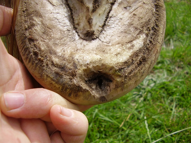
Seedy toe: a term commonly used for localized separation of hoof horn at the center of the toe.

Examples of hooves with separation at the center of the toe, where a crena would be located
Terminology varies, particularly around seedy toe and white line disease. Neither term should be treated as another name for the bony notch.
Sato and colleagues studied 326 front hooves from 163 Thoroughbred yearlings, examining the horses before their first shoeing. Greater notch depth and width were associated with higher grades of toe white line separation.

This finding suggests that local anatomy may influence susceptibility. It does not establish that separation creates the crena, or that every horse with a large crena will develop separation. Some hooves with deeper or wider notches had little or no corresponding abnormality.
The practical implication is to assess the horn at the toe carefully. A radiographic feature and an active hoof defect can coexist, but each needs its own interpretation.
Our White Line Disease Management Guide explains hoof wall separation, assessment, and management in greater detail.
What Should Owners and Farriers Do?
An incidental crena in a comfortable horse does not, by itself, establish a need for corrective treatment. Decisions should reflect the horse’s soundness, the condition of the hoof capsule, and any associated clinical findings.
Regular hoof care provides opportunities to identify changes. Watch for a persistent toe pocket, repeated debris accumulation, crumbly horn, or separation that is enlarging. These findings warrant professional assessment even if the horse remains sound. White line disease can be present before obvious lameness develops.
A veterinarian may recommend radiographs when separation is extensive, recurrent, or accompanied by pain. Imaging helps assess the internal structures and extent of a defect; interpretation should consider the examination and history. Merck Veterinary Manual
Practical management includes:
Maintain an appropriate farriery schedule. Base the interval on hoof growth, wear, and the individual horse.
Assess the whole foot. Our Hoof Balance in Horses guide explains why external appearance alone is insufficient.
Review toe mechanics when separation is present. Understanding Breakover and The Rolled Toe Horseshoe explain relevant mechanical principles.
Treat an identified horn defect appropriately. Removal of compromised horn and any necessary support should be planned by the farrier and veterinarian.
Investigate pain promptly. Lameness or recurrent abscesses should not automatically be attributed to a normal anatomical notch. Merck Veterinary Manual
Research has not established a particular trim or shoe as a reliable way to prevent crena formation. Hoof care should address the horse’s functional needs and any actual disease.
Frequently Asked Questions
1. What is a notched coffin bone?
It is a coffin bone with an indentation called the crena marginalis at the center of its lower toe border. This can be a normal anatomical variation.
2. Is every horse born with a crena?
No. Some horses lack a detectable notch. Foal research shows that it can first become radiographically visible during the weeks or months after birth.
3. Does a crena mean the coffin bone is diseased?
No. The finding alone does not diagnose disease. A veterinarian interprets its appearance alongside other imaging findings, clinical signs, and the horse’s history.
4. Do toe clips cause the crena?
The 2025 radiographic study found no significant association between crena presence and the shoeing groups examined, including shoes with toe clips.
5. Is the crena inherited?
Inheritance is a possible explanation for some anatomical differences, but it has not been established. Differences between breeds do not, by themselves, demonstrate a genetic mechanism.
6. Is a crena the same as seedy toe?
No. The crena concerns bone shape. Seedy toe describes a localized defect in hoof horn at the toe, although terminology varies among practitioners.
7. Does a larger crena guarantee white line separation?
No. Larger notches were associated with more severe separation in one Thoroughbred yearling study, but individual findings varied. Association does not predict every horse’s outcome.
8. Can I identify the crena by looking underneath the hoof?
You can inspect the horn and white line, but you cannot directly see the bone through an intact hoof. Imaging is needed to assess the notch itself.
9. Does a horse with a crena need special shoes?
The notch alone does not determine shoeing requirements. Decisions should consider comfort, hoof condition, workload, and any associated problem requiring support or altered mechanics.
10. When should a toe defect be assessed?
Arrange assessment for persistent or expanding separation, recurrent abscesses, or crumbling horn. Seek prompt veterinary advice for lameness or other signs of pain.
For further study, explore our Equine Anatomy Study Kit and Equine Lameness Study Kit.
References
Smallwood, J. E., Albright, S. M., Metcalf, M. R., Thrall, D. E., and Harrington, B. D. (1989). A xeroradiographic study of the developing equine foredigit and metacarpophalangeal region from birth to six months of age. Veterinary Radiology, 30, 98–110. DOI: 10.1111/j.1740-8261.1989.tb00753.x.
Szabó, L., and Nagy, A. (2025). Computed tomographic evaluation of protrusion of the superficial layer of the hoof wall and crena marginis solearis in non-lame horses. Equine Veterinary Journal, 57(S61), 34–35. Conference research abstract. DOI: 10.1111/evj.70061.
Ennsmann, L. H., and Licka, T. F. (2025). Association between radiographic equine distal phalanx characteristics and absence, presence and type of horseshoes. Frontiers in Veterinary Science, 12, 1598038. DOI: 10.3389/fvets.2025.1598038.
Sato, F., Otsuka, N., and Kuwano, A. (2021). Toe white line separation associated with the notch (crena marginalis) on the distal phalanges in Thoroughbred yearling horses. Journal of Veterinary Medical Science, 83(11), 1745–1749. DOI: 10.1292/jvms.21-0270.
Beasley, B. (2024). White Line Disease in Horses. Merck Veterinary Manual.
University of Minnesota Extension. Caring for Your Horse’s Hooves.


Comments