Introduction to the Equine Hock: Anatomy, Biomechanics, and Why It Matters
- Horse Education Online

- May 21
- 8 min read

The equine hock, also called the tarsus, is one of the most important structures in the horse’s hind limb. It plays a central role in movement, athletic performance, balance, and soundness. Whether a horse is sprinting down a racetrack, collecting in a dressage arena, navigating rough trail terrain, or simply walking through a pasture, the hock is heavily involved in every stride.
Although the hock is often compared to the human ankle, it is far more specialized. It is designed to withstand enormous forces while still allowing the horse to move efficiently and powerfully.

Because of this, the hock is also one of the most common locations for arthritis, strain, and performance-related lameness.
Understanding how the hock works can help horse owners better recognize early signs of discomfort, make informed decisions about hoof care and training, and appreciate just how much stress this region endures throughout a horse’s life.
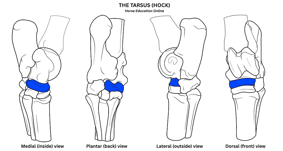
What Is the Equine Hock?
The hock is the large joint region located on the hind limb between:
The tibia above
The metatarsus, or cannon bone, below
From the side, the hock creates the angled appearance of the horse’s hind leg. The prominent point at the back of the hock is formed by the calcaneus, which acts as a major attachment point for powerful tendons.

Unlike a simple hinge joint, the hock is actually a complex system of multiple joints stacked together. These joints work in coordination to create movement, absorb concussion, and stabilize the hind limb during athletic activity. This combination of mobility and stability is what allows horses to generate tremendous forward power while still remaining balanced and coordinated.
Why the Hock Is So Important
Horse professionals often describe the hindquarters as the horse’s “engine,” and the hock is a major part of that engine.
The hock is responsible for:
Generating propulsion
Supporting collection
Absorbing impact
Stabilizing the hind limb
Storing and releasing elastic energy
Assisting with balance and turning
If the hock becomes painful or dysfunctional, the effects are often seen throughout the entire horse. Horses with hock pain may lose impulsion, struggle to engage the hindquarters, shorten their stride, or become resistant under saddle.
In many cases, subtle performance problems begin in the hock long before obvious lameness appears.
Bones of the Equine Hock
The equine hock consists of several small bones arranged in rows.
Proximal Row
The upper row contains:
Talus
Calcaneus
The calcaneus forms the visible “point” of the hock and acts as a lever arm for powerful tendon attachment.

Middle Bone
Central tarsal bone
Distal Row
1st and 2nd tarsals (fused)
3rd tarsal
4th tarsal

These bones are connected by multiple small joints, ligaments, and layers of cartilage that allow the hock to function smoothly under heavy stress.
While the individual bones are relatively small, together they create an extremely strong and efficient mechanical system.
The Four Main Joints of the Hock
The hock contains four primary joints, and a fifth secondary joint, each serving different functions.
1. Tibiotarsal Joint (Tarsocrural Joint)
This is the largest and most mobile joint in the hock.
It is responsible for approximately 90% of total hock movement and provides most of the flexion and extension seen during locomotion.
This joint is especially important for:
Jumping
Collection
Acceleration
Stride length
Athletic maneuvering

When a horse flexes the hock deeply during collected work or jumping effort, most of that movement occurs in the tibiotarsal joint.
Because this joint has significant motion, it also contains a large amount of synovial fluid and cartilage to reduce friction.
2. Proximal Intertarsal Joint
This joint sits between:
The talus and calcaneus above
The central tarsal bone below

It has limited movement compared to the tibiotarsal joint, but still contributes to flexibility and force transfer.
Its main role is helping distribute forces throughout the hock while maintaining stability.
3. Distal Intertarsal Joint
The distal intertarsal joint has very little motion.
Instead of functioning primarily as a movement joint, it acts more as a shock-transfer and stabilization structure.

Unfortunately, this joint is subjected to heavy compressive forces over time, making it a common location for osteoarthritis.
This joint is frequently involved in bone spavin, one of the most common forms of hock arthritis in horses.
4. Tarsometatarsal Joint
This joint connects the lower tarsal bones to the cannon bone.
Like the distal intertarsal joint, it has minimal movement and functions mainly as a weight-bearing support structure.

It acts almost like a rigid platform during propulsion, helping transfer force from the hindquarters into forward motion. This joint is also commonly affected by degenerative joint disease.
Talocalcaneal Joint
In addition to the four major hock joints commonly discussed, the equine hock also contains the talocalcaneal joint, sometimes referred to as the talocalcaneal articulation. This joint exists between the talus and calcaneus, the two large bones in the proximal row of the hock. Unlike the tibiotarsal joint, the talocalcaneal joint has very limited movement and functions primarily as a stabilizing structure. Its main role is helping transfer forces through the upper hock while maintaining alignment between the major tarsal bones during motion.

Strong ligaments surrounding the joint provide additional support, allowing the hock to withstand substantial biomechanical stress during propulsion and weight-bearing. Although it is not typically a primary source of clinical lameness, the talocalcaneal joint contributes to the overall stability and mechanical efficiency of the equine hock complex.
How the Hock Produces Power
One of the hock’s most important functions is generating propulsion.
When the horse pushes off the ground:
The hock extends forcefully
Tendons tighten
Muscles contract
Energy transfers through the limb
This creates the forward-driving power that propels the horse ahead.
The hock works closely with:
The pelvis
Gluteal muscles
Hamstrings
Stifle
Back muscles
Together, these structures create coordinated hindquarter movement.
A healthy hock allows the horse to:
Push from behind
Carry weight effectively
Develop impulsion
Move with athletic power
When hock pain develops, horses often begin moving “flat” or lose engagement behind.
The Hock as a Shock Absorber
Every time the hind hoof strikes the ground, concussion travels upward through the limb.
The hock helps absorb and redistribute these forces.
Several structures contribute to this function:
Articular cartilage
Synovial fluid
Ligaments
Tendons
Joint surfaces
Without this shock absorption system, stress on the upper limb and spine would increase dramatically.
This protective role is especially important in:
Jumping horses
Reining horses
Barrel horses
Racehorses
Horses working on hard footing
Repeated concussion over time can contribute to wear and degeneration within the lower hock joints.
Elastic Energy Storage
The hock also functions like a spring.
During movement:
Tendons stretch under load
Elastic energy is temporarily stored
That energy is released during push-off
This improves movement efficiency and reduces muscular fatigue.
The horse does not rely solely on muscle strength. Instead, tendons and ligaments help recycle energy with each stride.
This is one reason horses can move so efficiently over long distances.
Stability and Balance
Although the upper hock joint is mobile, the lower joints are comparatively rigid.
This rigidity is intentional.
The lower hock joints provide:
Stability
Weight-bearing support
Balance control
Resistance to twisting forces
These features are especially important during:
Turning
Lateral movements
Uneven terrain
Sliding stops
Quick acceleration
Too much motion in the lower joints would reduce stability and increase injury risk.
The Role of the Hock in Collection
Collection requires the horse to shift more weight onto the hindquarters.
To do this effectively, the horse must flex the hock more deeply.
In collected work:
The hindquarters lower
The hock compresses
The horse carries more weight behind
Balance shifts rearward
This allows for elevated, controlled movement.
Disciplines that heavily rely on hock engagement include:
Dressage
Reining
Cutting
Jumping
Eventing
However, increased collection also increases pressure within the hock joints. Over time,
repetitive compression can contribute to arthritis and degenerative change.
Tendons and Ligaments Supporting the Hock
The hock depends on strong soft tissue support structures.
Collateral Ligaments
These stabilize the hock from side-to-side motion and help prevent excessive twisting.

Long Plantar Ligament
This major ligament supports the back of the hock and contributes to stability during weight-bearing.

Calcanean Tendon Group
Often compared to the human Achilles tendon, this structure allows powerful extension of the hock during propulsion.
Together, these tissues help the hock withstand tremendous biomechanical stress.
Dorsal Ligament
The dorsal ligament, also called the talocentrodistometatarsal ligament, is an important stabilizing ligament on the front side of the hock. It connects the dorsal, or front, surface of the talus to the lower portions of the central tarsal bone, third tarsal bone, and cannon bone. Its main role is to help stabilize the stacked bones of the hock and support proper force transfer during weight-bearing and movement.

Why Hock Problems Are So Common
The hock is highly vulnerable to injury and degeneration because it experiences:
Repetitive stress
High compressive forces
Constant motion
Athletic loading
Concussion from hard ground
Even small imbalances in movement or hoof trimming can gradually alter how forces travel through the hock. Over time, this may contribute to inflammation, cartilage wear, and arthritis.
Hind limb lameness in horses is frequently attributed to the hock, stifle, or back, and distinguishing between these regions can be challenging even for experienced professionals. Read "Is it Hock, Stifle, or Back Pain? How to Tell the Difference" to learn more.
Common Hock Conditions
Bone Spavin
Bone spavin is osteoarthritis affecting the lower hock joints.
Common signs include:
Stiffness
Shortened stride
Difficulty engaging the hindquarters
Resistance under saddle
Reduced performance
It is especially common in athletic horses and older horses.
Thoroughpin
A thoroughpin is distension of the tendon sheath near the upper back portion of the hock.
It often appears as a soft swelling and may or may not cause lameness.
Capped Hock
Capped hock is swelling over the point of the hock, usually caused by trauma or repeated pressure.
It is often cosmetic but may occasionally become inflamed.
OCD Lesions
Osteochondritis dissecans (OCD) lesions are developmental orthopedic problems seen more commonly in young horses.
These lesions involve abnormal cartilage and bone development within the joint.
Hock Lameness Study Aids
Dive deeper into hock-related lameness by enrolling in the Equine Lameness Certification Program, and take advantage of the following study aids:
Early Signs of Hock Pain
Recognizing subtle signs early can help prevent more severe problems later.
Common warning signs include:
Shortened hind stride
Stiffness when first moving
Difficulty picking up leads
Resistance to collection
Reduced impulsion
Toe dragging
Frequent stumbling
Trouble holding a canter
Behavioral changes under saddle
Because hock discomfort may develop gradually, changes are often mistaken for laziness or aging.
Hoof Balance and the Hock
The hoof and hock are closely connected biomechanically.
Poor hoof balance can alter:
Limb loading
Breakover
Joint compression
Tendon strain
Improper trimming or shoeing may increase stress on the hock over time.
This is one reason veterinarians and farriers often work together when managing horses with hock pain.
Correct hoof balance can help improve comfort and reduce abnormal stress patterns throughout the hind limb.
For readers interested in hoof balance and lower limb biomechanics, Horse Education Online offers additional educational articles and study materials covering equine anatomy, lameness, and hoof care principles.
Q&A About the Equine Hock
What is the equine hock?
The equine hock, also called the tarsus, is the large joint complex in the horse’s hind limb between the tibia above and the cannon bone below. It helps generate power, absorb shock, stabilize the hind limb, and support athletic movement.
Is the horse’s hock the same as the human ankle?
The horse’s hock is often compared to the human ankle, but it is more specialized for high-force movement. It is built to handle propulsion, balance, concussion, turning, jumping, collection, and repeated athletic loading.
Which joint in the hock has the most movement?
The tibiotarsal joint, also called the tarsocrural joint, is the most mobile joint in the hock. It provides most of the flexion and extension used during movement, especially during jumping, acceleration, stride lengthening, and collection.
Why is the hock important for horse movement?
The hock is a major part of the horse’s hindquarter power system. It helps the horse push from behind, carry more weight on the hind end, develop impulsion, absorb concussion, and maintain balance during turns, transitions, and athletic work.
What are common signs of hock pain in horses?
Common signs of hock pain include stiffness, shortened hind stride, toe dragging, reduced impulsion, difficulty picking up leads, resistance to collection, trouble holding the canter, frequent stumbling, and behavioral changes under saddle.
What is bone spavin in horses?
Bone spavin is osteoarthritis affecting the lower hock joints. It is a common cause of hind limb stiffness, shortened stride, reduced performance, and difficulty engaging the hindquarters, especially in older or athletic horses.
Can hoof balance affect the hock?
Yes. Hoof balance can influence breakover, limb loading, tendon strain, and joint compression. Poor trimming or shoeing can increase abnormal stress through the hock over time, which is why veterinarians and farriers often work together when managing hock pain.


Comments