Equine Protozoal Myeloencephalitis (EPM): An Advanced Clinical and Management Perspective
- Horse Education Online
- 6 days ago
- 13 min read
Updated: 6 hours ago
Equine protozoal myeloencephalitis, or EPM, is one of the most important infectious neurologic diseases affecting horses in the Americas. What makes it clinically challenging is not just the parasite itself, but the way EPM can mimic other neurologic conditions, vary from horse to horse, and force decisions that depend on lesion location, stage of disease, and realistic management goals.
This article is best read as a deeper clinical overview rather than a first-stop owner guide. It looks beyond early signs to the bigger framework: parasite biology, host roles, why horses are aberrant hosts, why horse-to-horse spread does not occur, and how those facts shape diagnosis, prevention, and long-term case management. We recommend horse owners start with our article EPM in Horses: Early Signs, Diagnosis & Management.
Quick answer
EPM is a protozoal neurologic disease of the central nervous system, most commonly caused by Sarcocystis neurona, and less commonly by Neospora hughesi. Opossums are the definitive host for Sarcocystis neurona, while horses are aberrant or accidental hosts that develop disease but do not continue the parasite’s normal life cycle. That is why prevention is mainly about reducing environmental exposure rather than isolating affected horses.
Clinically, EPM matters because signs can be highly variable and overlap with other neurologic disorders. The advanced perspective is not just “recognize the signs,” but understand that diagnosis, prognosis, and management depend on the broader clinical context, not one finding in isolation.
Etiology and Parasite Biology
EPM is most commonly caused by Sarcocystis neurona, with Neospora hughesi representing a less common but clinically similar pathogen.
The life cycle of Sarcocystis neurona is central to understanding disease risk. Opossums act as the definitive host, shedding infectious sporocysts in their feces. Horses become exposed through ingestion of contaminated feed, water, or pasture. However, horses are considered aberrant hosts. This means they are not part of the parasite’s normal life cycle and typically do not develop the tissue cyst stage seen in intermediate hosts.
This aberrant host status has important implications. Unlike species that serve as natural intermediate hosts, horses do not effectively contribute to transmission. Instead, they represent a biological endpoint in which the parasite can invade the central nervous system but cannot complete its life cycle.
Types of Hosts in Parasitology
Host Type | Definition | Role in Parasite Life Cycle | Example (EPM Context) |
Definitive Host | The host in which the parasite reaches sexual maturity and reproduces sexually. | Essential for completing the parasite’s life cycle and producing infectious stages. | Opossum (sheds Sarcocystis neurona in feces) |
Intermediate Host | A host that harbors the parasite during a transitional or developmental stage, often involving asexual reproduction. | Required for development but not for sexual reproduction. | Typical wildlife hosts in the natural life cycle (not horses) |
Aberrant Host | A host that is not part of the parasite’s normal life cycle and in which development is abnormal or incomplete. | Parasite may cause disease but cannot complete its life cycle. | Horse (for Sarcocystis neurona) |
Paratenic (Transport) Host | A host in which the parasite survives without further development, remaining infective to the next host. | Not required for development but can help bridge transmission between hosts. | Some small animals may serve this role in certain parasite cycles |
Reservoir Host | A host that maintains the parasite in nature and serves as a source of infection for other species. | Critical for persistence of the parasite in the environment. | Wildlife species that harbor stages of Sarcocystis neurona |
Accidental Host | A host that becomes infected unintentionally and is not typically involved in transmission. | Infection is incidental and often does not contribute to spread. | Horses can also be described this way in EPM context |
How This Applies to EPM
Understanding these host categories clarifies why EPM behaves the way it does:
The opossum, as the definitive host, is central to environmental contamination.
The horse, as an aberrant or accidental host, develops disease but does not contribute to spreading it.
The parasite’s life cycle depends on other species, not horses, which is why horse-to-horse transmission does not occur.
Practical Insight
This classification is not just academic. It directly informs prevention strategies:
Because horses are aberrant hosts, controlling exposure is far more important than isolating affected horses.
Because opossums are definitive hosts, management practices should focus on limiting their access to feed and water sources.
Understanding the broader host system helps explain why exposure is common, but disease is relatively uncommon.

Risk factors and exposure pathways
Understanding exposure risk matters because EPM prevention is not mainly about isolating affected horses. It is about reducing the chance that horses ingest infective material in the first place.
Why exposure happens
Horses are exposed when feed, forage, pasture, or water becomes contaminated with infective sporocysts. In practical terms, that usually means wildlife access to places where horses eat and drink.
The most important point for owners is this: the horse does not need direct contact with another horse with EPM to be at risk. The problem is environmental contamination, not routine horse-to-horse spread.
Common risk pathways on real farms
Exposure pathway | Why it matters | Practical example |
Feed room contamination | Grain and bagged feeds attract wildlife | Opossums gaining access to open feed bins or spilled grain |
Hay contamination | Hay stored where wildlife can climb, nest, or defecate increases risk | Hay stacked in open sheds or poorly secured loft areas |
Water contamination | Shared troughs, open tanks, and poorly protected water sources can be exposed | Wildlife crossing around troughs at night |
Pasture contamination | Horses may graze near contaminated areas without obvious signs | Horses eating near fence lines, barns, or feed waste areas |
Attractants around the barn | Feed waste, pet food, garbage, and fallen fruit bring wildlife closer | Opossums repeatedly visiting the same barn zone |
Barn-level factors that may increase exposure pressure
Some environments are simply harder to control than others. Risk may be higher when barns have:
easy wildlife access to feed storage
chronic feed spillage
unsecured garbage or pet food
outdoor feeding areas that attract scavengers
water sources that are hard to protect or clean
wooded edges or structures that make wildlife traffic more likely
This does not mean every exposed horse develops EPM. It means exposure opportunity is part of the equation, and good management should focus on reducing it.
A practical prevention mindset
The goal is not to create a “sterile” farm. The goal is to reduce unnecessary contamination points.
Useful prevention habits include:
storing grain in sealed containers
cleaning up spilled feed quickly
limiting wildlife access to hay and concentrates
protecting water sources as much as possible
removing barn attractants such as garbage, pet food, or fallen fruit
watching for repeated wildlife activity around feeding areas
What this means clinically
This section matters because it changes how owners think about prevention. If you treat EPM like a simple contagious disease, you focus on the wrong control points. If you understand the exposure pathway, you focus on feed, forage, water, wildlife access, and environmental hygiene.
Exposure Versus Disease Development
Exposure and disease are not the same thing. Many horses may encounter Sarcocystis neurona without ever developing clinical EPM. This is one reason EPM diagnosis can be challenging: evidence of exposure does not automatically prove that the parasite is causing the horse’s current neurologic signs.
For EPM to develop, the parasite has to do more than enter the body. It must reach the central nervous system and cause damage significant enough to create clinical signs. That is why it helps to understand the basics of the equine nervous system before interpreting weakness, ataxia, muscle loss, or abnormal gait patterns.
This distinction matters clinically. A horse may test positive because of exposure but have neurologic signs caused by something else. Another horse may show a pattern that makes EPM more suspicious, especially if signs are asymmetric or difficult to explain as simple lameness. For owner-friendly early recognition, see EPM in Horses: Early Signs, Diagnosis & Management.
A practical way to think about it:
Situation | What it means |
Exposure without signs | The horse may have encountered the organism but is not clinically affected |
Positive test with neurologic signs | EPM may be possible, but the result still needs clinical interpretation |
Neurologic signs with unclear testing | Other neurologic diseases must stay on the differential list |
Confirmed or strongly suspected EPM | Treatment and management should be guided by the veterinarian’s full assessment |
The owner takeaway is simple: reduce exposure where possible, but do not assume exposure always becomes disease. EPM should be interpreted through the full case picture: history, neurologic exam, diagnostic testing, progression over time, and response to treatment. When signs are hard to localize, tools like the Equine Neuro Screen & Localization Assistant can help owners organize observations before speaking with their veterinarian.
For differential thinking, this is also where conditions such as equine herpesvirus matter. EPM is important, but it is not the only neurologic disease that can produce weakness, incoordination, or sudden performance changes.
Pathophysiology of Neurologic Disease
Once Sarcocystis neurona gains access to the horse, it can migrate into the central nervous system. The resulting inflammation leads to damage of neural tissues. This damage is often multifocal, meaning multiple areas of the brain or spinal cord may be affected simultaneously.
The clinical consequences depend on which neural pathways are involved. Damage to motor pathways can result in weakness or abnormal gait. Involvement of sensory pathways can lead to incoordination. Cranial nerve involvement produces deficits such as facial paralysis or difficulty swallowing.
A defining feature of EPM is that lesions are often asymmetric. This means neurologic deficits may be more pronounced on one side of the body, which can help differentiate EPM from some other neurologic conditions.

Reference: Review A review of Sarcocystis neurona and equine protozoal myeloencephalitis (EPM) J.P. Dubey a,∗ , D.S. Lindsay b,1 , W.J.A. Saville c , S.M. Reed d , D.E. Granstrome , C.A. Speerf,2
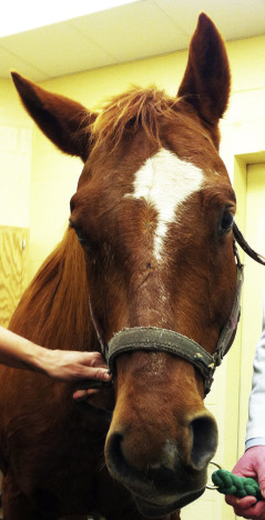
Clinical Presentation
EPM is often described as a disease of variability. Its clinical signs are determined by lesion location and severity, making no two cases identical. However, several categories of clinical findings are commonly recognized. The Neuro Screen & Localization Assistant can help with the identification of neurological diseases based on their symptoms.
Ataxia and Incoordination
Ataxia is one of the most frequently observed signs. Horses may appear unsteady, stumble, or have difficulty placing their limbs accurately. This incoordination is often asymmetric, which is a key clinical clue. Learn more about ataxia here.
Weakness and Abnormal Gait
Weakness may manifest as a shortened stride, dragging of the toes, or difficulty maintaining posture. In more advanced cases, horses may struggle to rise or may fall.
Muscle Atrophy
Muscle wasting can occur, often in a focal pattern. This may be particularly noticeable along the topline or in specific muscle groups and is associated with nerve damage.
Cranial Nerve Deficits
Involvement of cranial nerves can produce signs such as facial droop, difficulty chewing or swallowing, and abnormal eye or ear positioning.
Because these signs overlap with many other neurologic disorders, none of them are diagnostic on their own.


Examples of facial droop in horses
Diagnostic Challenges
Diagnosing EPM is one of the most complex aspects of managing the disease. A fundamental issue is that many healthy horses in endemic areas have antibodies against Sarcocystis neurona, indicating exposure rather than active infection.
The Role of the Neurologic Examination
A thorough neurologic examination is the cornerstone of diagnosis. This examination allows clinicians to localize lesions within the nervous system and determine whether findings are consistent with EPM.
Rule-Outs
Equally important is the exclusion of other conditions that can produce similar signs. These include:
Cervical vertebral stenotic myelopathy
Equine herpesvirus myeloencephalopathy
Trauma
Degenerative neurologic diseases
Failure to rule out these conditions can lead to misdiagnosis and inappropriate treatment.
Laboratory Testing
Antibody testing is commonly used, but interpretation must be cautious. Serum tests alone indicate exposure and are not sufficient to confirm disease.
More advanced diagnostic approaches focus on detecting intrathecal antibody production, meaning antibodies produced within the central nervous system. Paired serum and cerebrospinal fluid testing can improve diagnostic confidence.
Even with these tools, false positives and false negatives remain possible. For this reason, test results must always be interpreted in conjunction with clinical findings and other diagnostics.
Treatment Principles
The foundation of EPM treatment is antiprotozoal therapy. Peer reviewed consensus statements consistently identify this as the core approach to managing the disease.
Antiprotozoal Therapy
Antiprotozoal drugs target the causative organisms and aim to reduce or eliminate the parasite within the central nervous system. Early initiation of treatment is associated with improved outcomes.
Supportive Care
Supportive care is highly case-dependent and may include:
Anti-inflammatory strategies
Nutritional support
Controlled exercise programs
Environmental management to reduce stress
Each of these components plays a role in supporting recovery and minimizing further neurologic damage.
Rehabilitation
Once the horse is stabilized, controlled exercise and rehabilitation are often introduced. These programs aim to improve coordination, strength, and functional mobility. The timing and intensity of rehabilitation must be carefully managed to avoid exacerbating neurologic deficits.
Monitoring Response
Improvement in neurologic function may take weeks. Some horses show gradual progress, while others may plateau or require reassessment. Long-term monitoring is essential, as relapse or incomplete recovery can occur.
Prognosis and return-to-work expectations
One of the hardest parts of EPM management is that diagnosis and treatment are only part of the story. Owners also need a realistic view of what recovery may look like, how much function may return, and whether the horse is truly safe to ride again.
Prognosis is variable for a reason
EPM does not affect every horse the same way. Outcome depends on:
severity of neurologic deficits at presentation
how early the case is recognized
how the horse responds to treatment
which functions remain impaired after treatment
whether meaningful residual deficits remain
That is why it is not enough to ask, “Did the horse survive?” The more useful questions are:
How much neurologic function came back?
Is the horse safe?
Can the horse return to its previous job?
What improvement may look like
Some horses improve enough to return to useful work. Others improve but are left with mild residual deficits. Some remain unsafe for athletic use even if they look brighter and stronger than they did at diagnosis.
Clinical outcome | What it may mean in practice |
Marked improvement with minimal residual deficit | Some horses may return to prior work, depending on discipline and risk |
Partial improvement | Horse may become more comfortable but not fully reliable athletically |
Residual ataxia or weakness | Even mild persistent deficits may limit ridden safety |
Poor response or plateau | Long-term athletic return may be unrealistic |
Return to work is a safety decision, not just a fitness decision
A horse recovering from EPM should not be pushed back into work just because appetite improved or because treatment has finished. Return-to-work decisions should be based on:
repeat neurologic assessment
consistency of gait and coordination
ability to turn, back, and move safely
discipline demands
rider safety
environment and footing
A horse with residual neurologic impairment may still look “better” while remaining unsafe under saddle.
A more realistic way to counsel owners
For many horses, a better conversation is:
not “When can I compete again?”
but “What level of function is returning, and what is safe?”
That is especially important in horses expected to jump, work at speed, travel over uneven footing, or carry less experienced riders.
Practical owner expectations
Owners should prepare for the possibility that:
recovery may be slow
improvement may be incomplete
the horse may return to a lower level of work than before
follow-up reassessment matters more than optimism alone
A useful way to frame progress
The best signs are not just that the horse looks brighter. They are that the horse becomes:
more coordinated
more symmetrical
safer in turns and transitions
more reliable on repeat exams over time
That is a stronger basis for return-to-work planning than enthusiasm or appearance alone.
Prevention and Risk Reduction
Because EPM is associated with environmental exposure to infectious stages, prevention strategies focus on reducing contact with opossum feces and contaminated feed or water.
Feed and Storage Management
Store grain and feed in sealed containers
Clean up spilled feed promptly
Avoid leaving feed exposed in areas accessible to wildlife
Environmental Management
Limit opossum access to barns and feed rooms
Remove attractants such as garbage or pet food
Maintain clean water sources
Biosecurity Awareness
Understanding that exposure is common but disease is less frequent underscores the importance of management practices. Risk factor studies have shown that environmental and management conditions play a significant role in disease development.
When Immediate Veterinary Care Is Required
Certain clinical signs indicate the need for urgent veterinary evaluation:
Rapid progression of ataxia or weakness
Falling or inability to rise
Severe asymmetry in neurologic function
Difficulty swallowing or signs of choking
These signs suggest significant neurologic compromise and require prompt assessment to determine the underlying cause and initiate appropriate treatment.
Integrating Knowledge Into Practical Horsemanship
For horse owners and professionals, understanding EPM extends beyond recognizing clinical signs. It involves integrating knowledge of disease biology, risk factors, and management practices into daily care routines.
Recognizing subtle changes in gait, coordination, or behavior can lead to earlier detection. Maintaining consistent feeding and storage practices reduces exposure risk. Working closely with a veterinarian ensures that diagnostic and treatment decisions are based on a comprehensive evaluation rather than isolated findings.
Key Takeaways
EPM is a neurologic disease primarily caused by Sarcocystis neurona, with Neospora hughesi as a less common cause.
Clinical signs are variable and depend on lesion location within the central nervous system.
Diagnosis requires a combination of neurologic examination, laboratory testing, and exclusion of other diseases.
Serum antibodies indicate exposure, not necessarily active disease.
Antiprotozoal therapy is the foundation of treatment, supported by case specific care.
Prognosis varies, with many horses improving but some retaining residual deficits.
Prevention focuses on reducing exposure through environmental and feed management.
This article is intended for educational purposes and should not replace professional veterinary advice. For concerns about neurologic disease in horses, consultation with a qualified veterinarian is essential.
EPM Q&A
What is equine protozoal myeloencephalitis in horses?
Equine protozoal myeloencephalitis, often called EPM, is a neurologic disease that affects the horse’s central nervous system. It can involve the brain, spinal cord, or both, which is why signs may include weakness, ataxia, muscle loss, gait changes, or other neurologic abnormalities.
What causes equine protozoal myeloencephalitis?
EPM is most commonly associated with Sarcocystis neurona. A less common cause is Neospora hughesi. Horses are exposed when they ingest infective material from contaminated feed, hay, pasture, or water, most often connected to wildlife contamination.
Can horses spread EPM to each other?
No. EPM is not considered a horse-to-horse contagious disease. Horses are aberrant or dead-end hosts, meaning they can develop disease but do not continue the parasite’s normal life cycle or spread it directly to other horses.
Why is EPM diagnosis difficult?
EPM diagnosis is difficult because exposure does not always mean disease, and neurologic signs can overlap with other conditions. A positive test result must be interpreted with the horse’s history, neurologic exam, clinical signs, diagnostic testing, and differential diagnosis.
What diseases can look like EPM in horses?
Other neurologic conditions can resemble EPM, including cervical vertebral stenotic myelopathy, equine herpesvirus neurologic disease, trauma, equine degenerative myeloencephalopathy, and other causes of weakness or ataxia. This is why EPM should not be diagnosed from one sign or one test alone.
What is the prognosis for horses with EPM?
The prognosis for EPM varies. Some horses improve significantly with treatment, while others retain neurologic deficits. Outcome depends on severity, how early the disease is recognized, response to treatment, lesion location, and whether residual ataxia or weakness remains.
Can a horse return to work after EPM?
Some horses can return to work after EPM, but return-to-work decisions should be based on neurologic safety, not just whether the horse looks brighter or has finished treatment. Repeat assessment, coordination, gait consistency, discipline demands, and rider safety all matter.


