Horse Skeleton Anatomy Explained: Major Bones, Diagram, and Function
- Horse Education Online

- Mar 9
- 17 min read
The horse skeleton is the framework that gives your horse shape, strength, and structure. It is also the “map” vets, therapists, and trainers are thinking about when they talk about joints, lameness, posture, and movement.
In this guide, you’ll learn the major bone regions, what each region does, and why skeleton anatomy matters for everyday horse care. We’ll keep it owner friendly, with the names you actually need and the practical reasons they matter.
If you want to see every bone in 3D while you read, open the Interactive Horse Skeleton tool in another tab. It pairs perfectly with this article. You can also explore how bones connect across systems using the Interactive Horse Anatomy hub.
One important difference from humans is how much the horse skeleton is built for forward motion and weight bearing. Horses do not have collarbones, their spine is designed to support a rider, and their limbs are engineered for efficient stride and shock absorption. That is why basic horse skeleton anatomy shows up in everything from conformation talk to soundness discussions.
What the horse skeleton does
The skeleton is not just a collection of bones. It is a working structure that supports the horse’s body, protects important organs, and creates the lever system that makes movement possible.
Support and weight bearing
A horse carries a large percentage of body weight on the front end, and the skeleton is built to handle that load repeatedly, day after day. Bones give the body a strong frame, while joints and soft tissues let the frame move without falling apart.
When you think about soundness, you are often thinking about how well this structure is handling stress over time. If you want a step by step way to connect anatomy to real world soundness checks, the comprehensive guide to equine lameness assessment explains how vets and experienced evaluators work through the body logically.
Protection of the brain, chest, and internal organs
Some bones are built to move, and others are built to protect. The skull protects the brain and supports the structures of breathing and chewing. The rib cage protects the heart and lungs. The spine protects the spinal cord and also acts like a bridge between the front and hind end.
This is one reason anatomy matters in emergencies. Knowing what is under a swelling, a wound, or a painful area helps you describe what you see clearly and respond faster.
Movement and athletic function
Bones are the rigid “bars” in the lever system of movement. Joints are the hinges. Muscles provide the pulling power. Tendons and ligaments guide motion and store energy.
That is why the skeleton is not taught in isolation. If you want to see how muscles attach and work over the skeleton in motion, pair this section with the Interactive Horse Muscles tool. It makes the “why” behind bone shapes and joint angles much easier to understand.
In the next sections, we’ll break the skeleton into big regions first, so you have a clear mental map before we name individual bones.
Axial vs appendicular skeleton in horses
A simple way to understand horse skeleton anatomy is to split it into two big parts. This helps you “place” what a vet, trainer, or anatomy diagram is talking about before you memorise bone names.
Think of it like this: the axial skeleton is the horse’s central frame (head, spine, rib cage), and the appendicular skeleton is the limb system (forelimbs and hindlimbs) that attaches to that frame.

If you want to see both systems in one place while you read, the Interactive Horse Anatomy hub makes this click quickly. It also pairs well with the equine nervous system guide because nerves run along the spine and into the limbs, which matters when pain patterns feel confusing.
Axial skeleton
The axial skeleton includes the skull, vertebral column (neck through tail), ribs, and sternum. Its main jobs are protection and support: it protects the brain and spinal cord, shields the heart and lungs, and provides the core structure the horse’s movement is built around.
Owner example: if a horse is sore through the back or guarded around the girth, the issue might involve structures supported by the axial skeleton (spine, ribs, sternum), even if the “problem” shows up as tension or short stride.
Appendicular skeleton
The appendicular skeleton includes the bones of the forelimbs and hindlimbs. These are the “engine and suspension” parts of the horse. Limbs are built for stride efficiency, shock absorption, and propulsion, with long bones acting as levers and joints acting like hinges.
Owner example: most obvious lameness you can see at the walk and trot is coming from the appendicular system, but the limb does not work alone. It is attached to the axial system through muscle slings and the pelvis.
Quick memory tip
If you can point to the horse’s spine and rib cage, you’re in axial territory. If you’re anywhere below the shoulder or hip into the leg, you’re in appendicular territory.
System | What it includes | Why owners care |
Axial | Skull, spine, ribs, sternum | Protects organs, supports posture, influences back comfort |
Appendicular | Forelimbs and hindlimbs | Drives movement, takes impact, where most visible lameness shows |
The skull and head bones

You do not need to memorise every tiny head bone to understand what matters. For owners, the key is knowing what the skull does for chewing, breathing, and protection, and how to describe locations clearly when something looks wrong.
If you want to “locate” head structures quickly, open the Interactive Horse Anatomy hub and use it like a map. For practical health context, the head also shows up in basic exams like gum colour and pulse checks, so keeping the horse’s vital signs guide handy is surprisingly useful.
The big structures to know
The skull protects the brain and forms the framework for the sinuses, nasal passages, and teeth. The mandible is the lower jaw and is central to chewing. The orbit is the bony socket that protects the eye. The nasal region shapes airflow and houses important sinus space.
Why teeth matter in a skeleton discussion

Teeth are not just “dental.” They are part of skull function. Chewing efficiency affects weight, behaviour under saddle, and sometimes even performance. When a horse is quidding, dropping feed, or resisting the bit, you are often looking at a head and jaw function problem first, not a “training” problem.
Owner relevant “where it matters” examples
If you see one sided nasal discharge, facial swelling, or foul odour, the nasal and sinus spaces become important to describe accurately. If a horse suddenly resists bridling or is head shy, jaw discomfort, tooth issues, or facial pain may be part of the story.
If you ever need to take a pulse quickly, most owners use the facial artery as a landmark. That ties directly into head anatomy and basic assessment, and this guide makes it simple to check and interpret: Average heart rate for a horse, what’s normal and how to check it.
Simple tip for better notes
When logging a head issue, write the side and the exact region. “Left cheek swelling” is more useful than “face swollen.” “Right nostril discharge, thick” is more useful than “runny nose.”
The spine of the horse

The spine is one of the most important regions in equine skeleton anatomy because it connects the entire horse. It supports posture, transmits force between the hind end and front end, and protects the spinal cord.
A helpful way to understand the spine is to think in regions rather than individual vertebrae. If you want a visual that makes posture and topline mechanics easier, pair this section with the Interactive Horse Muscles tool. For how the spine and pelvis connect into hind end movement, the equine hip joint deep dive is the best companion on your site.
Cervical vertebrae and neck support
Most horses have 7 cervical vertebrae. This region supports the head and allows the neck to flex, bend, and balance. Neck posture affects how a horse carries themselves, how they use their back, and how much strain ends up on certain soft tissues.
Practical example: a horse that braces the neck and avoids bending often looks “stiff,” but the whole body chain can be involved, including the back and pelvis.
Thoracic vertebrae and rib attachment
Most horses have 18 thoracic vertebrae. This region anchors the ribs and forms the framework for the withers area. It is also where saddle fit and rider weight become very relevant because the rib cage and thoracic spine are the “platform” under the saddle.
Practical example: a saddle that pinches at the withers or restricts shoulder movement can cause compensation patterns that look like lameness.
Lumbar vertebrae and the back
Most horses have 6 lumbar vertebrae, though there can be variation. The lumbar back is built more for strength and force transfer than extreme flexibility. This region matters for topline comfort, engagement, and how well the horse can round and carry.
Sacrum and connection to the pelvis

The sacrum is typically 5 fused vertebrae in adult horses. It forms the bridge between spine and pelvis. This is a major force transfer zone in propulsion, especially in athletic work.
Owner takeaway: when people say “hind end power,” they are describing a chain that runs through pelvis, hip, and sacral connection.
Tail vertebrae
The tail (coccygeal vertebrae) varies widely, often around 15 to 21 vertebrae. It is less about riding function and more about balance, communication, and muscle attachment.
Quick reference table
Counts can vary slightly by horse and by how sources count certain transitional vertebrae, but this gives a useful working map.
Spine region | Typical count | Main job (plain language) |
Cervical (neck) | 7 | Head and neck balance, steering, posture |
Thoracic (with ribs) | 18 | Rib support, withers structure, saddle platform |
Lumbar (back) | 6 | Strength and force transfer, back comfort |
Sacrum (fused) | 5 | Connects spine to pelvis for propulsion |
Tail | 15–21 | Balance, communication, muscle attachment |
Simple tip for owners
When you’re trying to describe back soreness, write where and what changed. For example: “flinch when grooming behind the withers,” or “tight when saddling at mid back,” or “short stride after sitting trot.” That kind of note is much easier for a professional to interpret than “back hurts.”
The rib cage and sternum
The rib cage is the protective “barrel” around the heart and lungs. Most horses have 18 pairs of ribs, attached along the thoracic spine. The ribs and spine together create the stable structure the horse breathes with and carries a rider on.
The sternum sits on the underside of the chest and helps support the chest floor. You do not usually think about the sternum until a horse is sore through the girth area, tight in the chest, or uncomfortable with saddle fit and breathing mechanics.
If you want to connect rib cage function to real, owner-level checks, pair this section with the horse’s vital signs guide. When breathing looks different, your first step is often respiration rate and effort, not guessing what bone is involved. For the broader “what counts as concerning” context, this early signs guide is a solid companion.
Why this region matters in breathing
The ribs move with each breath. If a horse is breathing harder at rest, showing heave-like effort, or guarding their chest, the rib cage is part of the story even when the primary cause is not “the ribs.”
Owner example: a horse with pain, fever, or respiratory illness may stand in a guarded posture and breathe differently. Your notes are more helpful if they describe effort and rate rather than only saying “breathing weird.”
Why this region matters in saddle fit and comfort
The saddle sits over the thoracic region, which includes the rib cage and thoracic spine. Poor saddle fit can create protective tension through the chest and back, and that can change how the horse moves.

Simple tip: if your horse looks short in front or reluctant to lift the back, take a quick note on “when it happens.” For example, only when saddled, only after 10 minutes, or only in one direction. Timing helps professionals separate training issues from discomfort patterns.
Quick reference
Structure | Plain-language job | Owner clue it may be involved |
Ribs | Protect heart and lungs, support breathing mechanics | Changes in breathing effort, guarding, girth sensitivity |
Sternum | Supports the chest floor | Discomfort at girth area, chest tightness, posture changes |
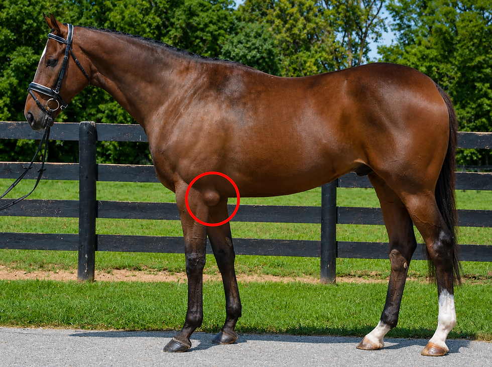
The forelimb skeleton

A lot of “where is it hurting” conversations start in the front end because the forelimbs handle a large share of weight bearing. The forelimb is built like a shock-absorbing pillar that supports the body and manages impact with every stride.
If you want the clearest clinical bridge from “bone names” to “how vets localize pain,” link this section to the comprehensive lameness assessment guide. For two common owner search topics that live in this region, the splint bone injuries overview and fetlock joint anatomy guide are perfect next clicks.
Shoulder and upper forelimb
The scapula (shoulder blade) and humerus form the upper limb. One key horse fact is that horses do not have a collarbone. Instead, the forelimb is attached to the body by a muscular “sling,” which helps absorb shock and allows efficient forward movement.
Owner example: when someone says a horse is “short striding in front,” it might not be the lower limb at all. It could be shoulder motion, muscle tension, or pain higher up that changes how the leg swings.
Radius, ulna, and the forearm
Below the humerus you have the radius and ulna. In horses, the ulna is reduced compared with humans, which supports the horse’s design goal: stable forward motion rather than rotating the forearm like we do.
A useful way to think about it is stability over versatility. Horses trade rotation for efficient stride and strength.
Knee and cannon region
The horse’s “knee” is the carpus. Below it is the cannon bone (metacarpal) and the splint bones alongside it. This area matters because it is a frequent site of swelling, knocks, and overuse changes.
If you ever see a new bump or sensitivity along the inside of the cannon region, it is worth reading the splint bone injuries guide so you can describe what you are seeing clearly and decide how urgent it is.
Lower limb bones
Below the cannon region, the key bones owners hear about are the pastern bones and the coffin bone, with the fetlock joint acting as a major motion and load region in between.
You do not need every small landmark, but knowing where the fetlock sits in the limb helps you follow lameness conversations quickly. The most owner-friendly deep dive is the anatomy of the fetlock joint.
Simple “where am I” table
Area owners point to | Anatomy term you will hear | Why it matters |
Shoulder | Scapula and humerus | Stride reach and front-end comfort |
Knee | Carpus | High-motion joint, common swelling site |
Cannon | Cannon bone and splints | Concussion, splint issues, tendon region |
Fetlock | Fetlock joint | Major load and motion, common lameness focus |
The hindlimb skeleton

If the forelimbs are the “front suspension,” the hindlimbs are the “engine.” The hind end is built for propulsion and power, pushing the body forward and helping the horse sit, jump, and accelerate.
To connect hindlimb anatomy to how soreness shows up in real movement, the best bridge is again the lameness assessment guide. For the core anatomy behind hind end power, the equine hip joint deep dive is your strongest internal link from this section.
Pelvis and hip region
The pelvis is the bony platform the hindlimb attaches to. It is built for force transfer. The hip region matters because it connects the spine to the hindlimb and influences stride length, stability, and how the horse uses the back.
Owner example: when a horse looks “not tracking up” or seems weaker behind, the issue might be anywhere in the hind end chain. Clean notes on when it appears (circles, transitions, hills, after rest) help professionals narrow the possibilities.
Stifle and gaskin region
The femur is the big upper hindlimb bone. The stifle is the joint system involving the femur, tibia, and patella. This area is central to “push” and also shows up in common issues like intermittent locking.
You do not need to diagnose stifle issues from a blog post, but it helps to know that the stifle is not a small joint. It is a major hinge and stabilizer that affects how the whole hindlimb loads.
Hock and lower hindlimb
The hock is the tarsus, and it is a key joint for propulsion and shock handling. Below it, the hindlimb continues with a cannon region and lower limb bones that mirror the forelimb in many ways.
Owner tip: when you write notes about hind end soreness, include whether the issue is worse on circles, worse at the start of work, or worse after work. Those timing details often matter more than the exact word you use for the location.
Quick comparison table
Limb system | Main job | What owners often notice first |
Forelimb | Weight bearing and shock absorption | Short stride in front, uneven landing, knee or cannon swelling |
Hindlimb | Propulsion and power | Reduced push, difficulty engaging, uneven tracking up |
Key joints that connect the skeleton
Bones do not work alone. The real “action points” are the joints, because that is where movement happens and where many common injuries and sore patterns show up. Owners also tend to describe problems by joint location, even if the underlying issue is bone, ligament, tendon, or soft tissue.
If you want to visualise how bones and muscles cross each joint, open the Interactive Horse Muscles tool while you read. It makes it much easier to understand why one sore area can change movement somewhere else.
Forelimb joints owners hear about most
The shoulder and elbow affect reach and how the leg swings forward. The carpus is the horse’s “knee,” and it is a common site of swelling and stiffness. The fetlock is a high motion, high load joint that works like a spring in stride.
If you want a clear, owner friendly explanation of what the fetlock does and why it is so important in soundness, use The Anatomy of the Fetlock Joint as the deeper companion section.
Hindlimb joints owners hear about most
The hip connects the hindlimb to the pelvis and influences stability and power. The stifle is a major hinge that affects how the horse engages and “pushes.” The hock is a key joint for propulsion and shock handling, and it often shows up in performance and stiffness conversations.
For a practical, detailed look at the hip region and how it supports hind end function, the best deep dive on your site is The Equine Hip Joint: An Advanced Deep Dive.
Quick joint language table
What owners say | Anatomy term | Why it matters |
Knee | Carpus | Common swelling and high motion joint |
Ankle | Hock (tarsus) | Propulsion and power, frequent stiffness site |
Pastern area | Pastern joints | Load transfer in the lower limb |
Fetlock | Fetlock joint | Spring and shock absorption, high load |
Tip for better vet communication: write the side and the joint. “Left front fetlock filling after work” is far more useful than “leg swollen.”
How the horse skeleton supports movement
A good way to think about movement is “support plus efficiency.” The skeleton supports weight, but it is also shaped to make forward motion easier and less costly.
If you want the best bridge between anatomy and what you see at the walk and trot, link this section to the comprehensive guide to equine lameness assessment. It helps readers connect bone and joint concepts to real world gait changes.
Weight bearing and shock absorption
Every stride is a controlled impact. The forelimbs tend to handle more weight bearing, while the hindlimbs contribute more propulsion. Joints, bone angles, and soft tissues work together to absorb force and prevent overload.
This is why “tiny” changes can matter. If a horse lands unevenly, loads one side more, or starts guarding a joint, the skeleton and soft tissues adapt. Those adaptations can become compensation patterns that look like stiffness, poor performance, or recurring soreness.
Leverage and stride length
Long bones act like levers. Joint angles determine how those levers move. This is one reason the same workload can feel easy for one horse and hard for another, even when both are fit.
A simple owner example is a horse that starts “shortening” in one direction on circles. That can be a joint comfort issue, a balance issue, or a compensation pattern. Your best clue is consistency and context: when it happens, which direction, and whether it improves with warm up.
Propulsion and the hind end “engine”
The hindlimb is built to push. The pelvis, hip region, stifle, and hock create a chain that drives the body forward. When that chain is not working smoothly, owners often notice reduced push, difficulty engaging, or trouble with transitions and hills.
If you want a concrete example of how limb imbalance and loading patterns can influence movement and soundness, Understanding High Low Syndrome in Horses is a strong internal bridge here because it explains how asymmetry shows up in real horses.
Quick movement notes that add value to your log
When you are writing movement observations, add one sentence that answers “when and where.” For example: worse on circles, worse after rest, improves after 10 minutes, or worse going downhill. Those small details often matter more than using the perfect anatomy word.
Why horse owners should understand basic skeleton anatomy
You do not need to become an anatomy expert. You just need enough horse bone anatomy knowledge to communicate clearly, notice changes earlier, and understand what professionals are telling you.
This is also where the article becomes useful beyond curiosity. Basic skeleton knowledge makes ownership easier.
It helps you describe problems accurately
When you can name the general region and joint, your notes become clearer. That improves vet calls, bodywork sessions, and training conversations. It also makes your own timeline more reliable because you are not guessing later what “front leg” meant.
A simple habit that helps is using the same structure in your notes every time: limb and side, region, what changed, and when it started.
It helps you understand imaging and vet explanations
Radiographs, ultrasound images, and exam notes make more sense when you can visualise the skeleton map. Even if you do not know every landmark, understanding the big bones and joints helps you follow the plan, ask better questions, and track progress.
It improves your learning speed across the whole site
Once you understand the skeleton regions, other topics become easier. Joints, muscles, nerves, and lameness discussions stop feeling like random terms and start feeling connected.
If you want to go deeper in a structured way, this is a natural place to explore the learning paths in membership, the step by step tracks in equine certifications, and the reference options in books and equine study materials.
Practical owner takeaway
Skeleton basics are not about memorising. They are about having a clear map. When you have the map, you spot changes sooner, communicate better, and make faster, calmer decisions.
Frequently asked questions about horse skeleton anatomy
How many bones does a horse have
Adult horses are often described as having around 200 bones, but the exact number can vary slightly depending on the source and how certain small bones are counted. Foals also have different counts because some bones fuse as the horse matures.
If you want a reliable way to learn the bones without worrying about exact totals, exploring the structures directly in the Interactive Horse Skeleton tool is more useful than memorising a single number.
What is the biggest bone in a horse
The femur (upper hindlimb bone) is commonly referenced as one of the largest and strongest bones in the horse, built to handle high loads during propulsion. The pelvis is also a major bony structure involved in force transfer.
To see how that region fits together in context, the equine hip joint deep dive is the best companion read.
Do horses have a collarbone
No. Horses do not have a collarbone. Their forelimb is attached to the body through a muscular sling rather than a bony connection, which helps with shock absorption and efficient forward movement.
If you want to see how that muscle sling relates to shoulder motion, use the Interactive Horse Muscles tool alongside the skeleton view.
What is the difference between the forelimb and hindlimb skeleton
The forelimbs are more focused on weight bearing and shock absorption, while the hindlimbs are more focused on propulsion and power. Both have similar “lower limb” patterns, but the pelvis and hip region make the hind end structurally different.
For a practical “how this shows up in movement” link, your lameness assessment guide helps readers connect anatomy to what they see at the trot.
What bone structures matter most in lameness exams
Vets localise lameness by combining observation, palpation, flexion tests, and sometimes diagnostic imaging. The major joints and the structures around them matter most, especially fetlock, hock, stifle, and the hoof region.
A strong next step for readers who want to understand that process is this comprehensive lameness assessment guide, because it explains the logic behind the exam in plain language.
Why is the horse spine different from a human spine
The horse spine is built to support a large body on four limbs while transmitting force between the hind end and front end. It must also support a rider and saddle without losing stability. It is less about twisting and more about strength, posture, and efficient force transfer.
If readers want to understand how nerves relate to the spine and why pain can “show up” in surprising areas, the equine nervous system guide is the best bridge.
What is the cannon bone
The cannon bone is the long bone between the knee (carpus) or hock and the fetlock. It is a major weight-bearing structure and a key area for tendons and ligaments.
If a reader is dealing with bumps or soreness along the cannon region, your splint bone injuries overview is a relevant internal link because splints run alongside the cannon bone.
What is the difference between the carpus and the hock
The carpus is the horse’s front “knee.” The hock is the hind “ankle” region, also called the tarsus. They are both complex joint areas, but they sit in different limbs and serve different roles in movement.
To understand how these joints fit into athletic function, the Interactive Horse Muscles tool makes the clearest visual connection.
Conclusion
The easiest way to learn horse skeleton anatomy is to start with the big regions, then connect each region to what it does. When you understand function, names become easier, movement makes more sense, and conversations about soundness feel far less confusing.
If you want the fastest “learn by exploring” method, use the Interactive Horse Skeleton tool to click through each bone and region. Then, if you want to build deeper knowledge with guided learning paths and structured resources, explore membership, equine certifications, and the reference library in equine study materials.


Comments