Laminitis in Horses: Causes, Signs, and Emergency Treatment
- Horse Education Online

- May 4
- 6 min read
Laminitis is a painful condition where the attachment between the hoof wall and the coffin bone becomes damaged. It is most commonly caused by high insulin levels, severe illness, or excessive weight bearing on one limb. Acute laminitis is an emergency that requires immediate veterinary care. Early treatment, especially continuous cooling of the feet, pain control, and proper support, can reduce damage. Long-term management depends on identifying and controlling the underlying cause.
What Is Laminitis?
Laminitis affects the lamellae inside the hoof. These are thousands of tiny structures that attach the hoof wall to the coffin bone.
When these structures are damaged, the bond between the hoof wall and coffin bone weakens. This can lead to instability, severe pain, and in advanced cases, rotation or sinking of the coffin bone.
Laminitis is typically described in two stages:
Acute laminitis: sudden onset of signs
Chronic laminitis: signs lasting longer than one week
Early recognition during the acute stage is critical to prevent permanent damage.
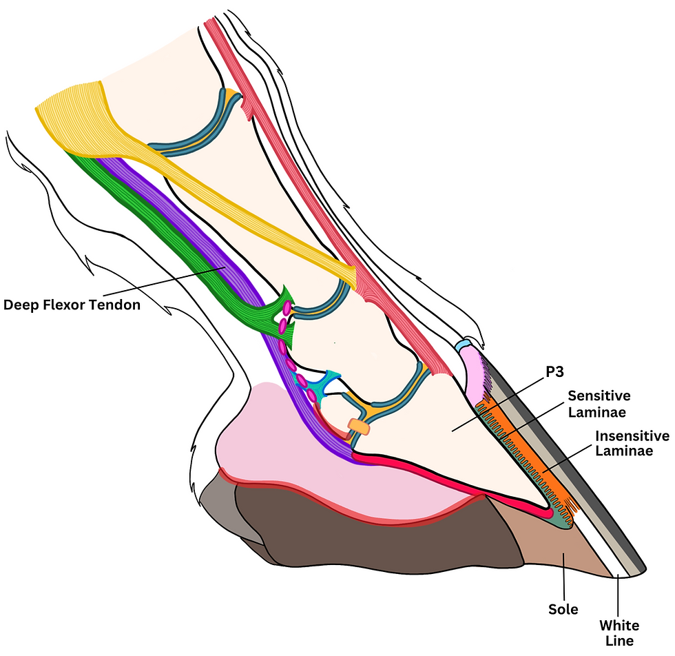
Anatomy Involved
To understand laminitis, we need to know what’s inside a horse’s hoof. Take a look at the drawing below and familiarize yourself with the structures of the horse’s hoof that are involved in laminitis. This will help you better understand the rest of the information contained in this article.
Coffin Bone: The coffin bone is the last bone in the horse’s leg, and the one closest to the ground.
Laminae: Think of the laminae as velcro: one side of the velcro is attached to the coffin bone. We call it the “sensitive laminae”. The other side of the Velcro is attached to the hoof wall. We call it the “insensitive laminae”.
These two layers interlock like Velcro and keep the hoof wall and the coffin bone joined together.
Hoof Wall: The hoof wall is a thick layer of dry, hard, and non-sensitive cells.
We call these cells “keratinized” or “cornified” cells, and they are the same type of cells that make up our fingernails and hair.
The purpose of the hoof wall is to protect the sensitive structures contained inside.
White Line: The white line is a continuation of the laminae. We discussed how the purpose of the laminae is to keep the hoof wall and the coffin bone attached. As the hoof wall grows down toward the ground, the “old” laminae grow past the bottom edge of the coffin bone.
Once the laminae grow past the bottom edge of the coffin bone, we begin calling it the “white line”. This is now the point of attachment between the hoof wall and the horse’s sole.
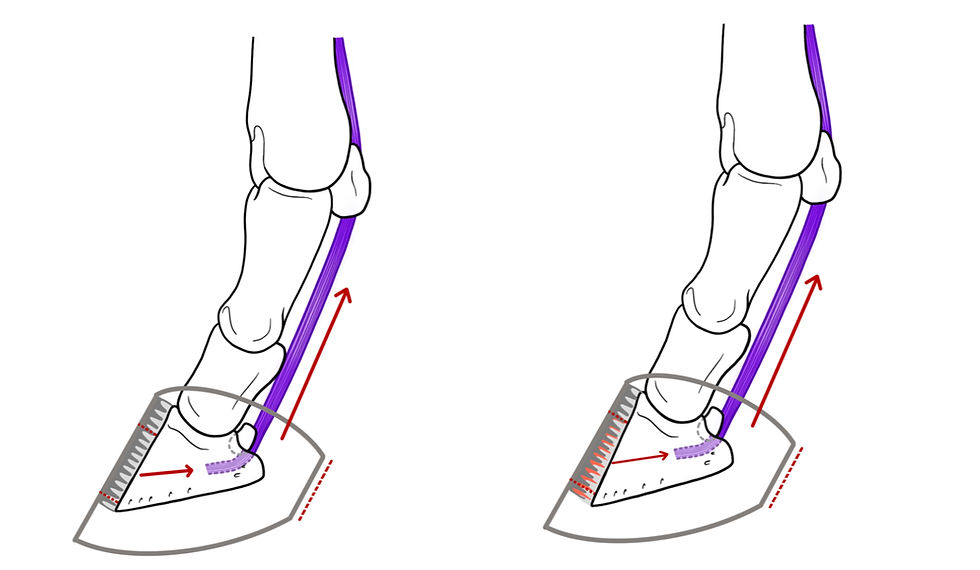
Why Laminitis Is So Painful
The lamellae support the horse’s entire body weight. When they fail, the hoof loses its ability to stabilize the coffin bone.
At the same time, the deep digital flexor tendon continues to pull on the bone. This creates mechanical stress that worsens the damage.
This combination leads to:
Structural failure inside the hoof
Increasing separation of tissues
Severe pain
Horses often show outward signs only after internal damage has already occurred.
To understand what laminitis feels like to the horse, think of a bruised nail.
Have you ever slammed your finger in a door, hit it with something heavy, or dropped a weight on your toe? That pressure under the nail can be intense and constant. This is similar to what happens inside a laminitic hoof.

The damaged laminae create pressure within the rigid hoof capsule. Unlike other parts of the body, the hoof cannot expand to relieve that pressure.
If you have experienced a bruised nail, you have felt a small fraction of the pain.
The difference is that you can protect your finger or toe. A horse cannot.
A horse must stand and bear weight on its feet, placing pressure on already damaged structures with every step.
Main Causes of Acute Laminitis
Endocrinopathic Laminitis
This is now the most common form.
It is linked to high insulin levels and commonly affects horses that:
Are overweight
Have PPID
Consume high sugar grass or feed
In this form, insulin directly damages the lamellae. It is not primarily an inflammatory condition. Read The Basics of Equine Nutrition – An Introductory Guide to understand the building blocks of a healthy equine diet and how nutrition directly impacts metabolic health.
Reference Study: Grenager NS. Endocrinopathic Laminitis. Vet Clin North Am Equine Pract. 2021 Dec;37(3):619-638. doi: 10.1016/j.cveq.2021.08.001. Epub 2021 Oct 19. PMID: 34674908.
Sepsis-Associated Laminitis
This form develops after severe illness such as:
Colic
Severe diarrhea
Retained placenta
Pneumonia or systemic infection
Inflammatory processes in the body contribute to lamellar damage.
Reference Study: Leise BS, Fugler LA. Laminitis Updates: Sepsis/Systemic Inflammatory Response Syndrome-Associated Laminitis. Vet Clin North Am Equine Pract. 2021 Dec;37(3):639-656. doi: 10.1016/j.cveq.2021.08.003. PMID: 34782098.
Supporting Limb Laminitis
This occurs when a horse avoids weight bearing on an injured limb.
The opposite limb becomes overloaded and can develop laminitis due to excessive stress.
Reference Study: van Eps A, Engiles J, Galantino-Homer H. Supporting Limb Laminitis. Vet Clin North Am Equine Pract. 2021 Dec;37(3):657-668. doi: 10.1016/j.cveq.2021.08.002. Epub 2021 Oct 19. PMID: 34674914.
Early Signs of Laminitis
Early signs can be subtle:
Strong digital pulse
Warm hooves
Reluctance to move
Short or stiff stride
Weight shifting
As the condition worsens:
The horse may lean back to relieve pressure
Movement becomes increasingly painful
The horse may lie down more often
Visible lameness often appears after internal damage has already occurred. To learn more about how subtle nutritional changes can affect your horse's movement and comfort, visit our equine lameness assessment guide.

What Is Happening Inside the Hoof?
The connection between the hoof wall and coffin bone begins to fail.
As this happens:
The coffin bone loses support
Tendon forces continue to pull on it
Damage progresses
This can lead to:
Rotation of the coffin bone
Sinking within the hoof capsule
Permanent structural changes
The longer the process continues, the more severe the damage becomes.

Emergency Treatment for Acute Laminitis
Immediate veterinary care is essential.
Continuous Cooling
Continuous cooling of the lower limbs and hooves is strongly supported by research.
This involves:
Keeping the feet in ice water
Maintaining cooling for extended periods
Early application can reduce tissue damage.
Pain Management
Laminitis causes severe pain.
Veterinary-prescribed medications are necessary to:
Improve comfort
Reduce stress
Prevent complications
Mechanical Support
There are many different ways to support a laminitic hoof, and no single method works for every horse. Options may include heart bar shoes, glue-on or screwed-in clogs, specialized therapeutic shoes, hoof casts, pads, or supportive boots. In some cases, simple solutions such as deep bedding or carefully applied frog support can also be effective.
Each of these applications works by redistributing weight, reducing strain on the damaged lamellae, and improving comfort. The goal is always to support the internal structures of the hoof while minimizing further mechanical stress.
The correct choice will vary from horse to horse. Laminitis does not affect every foot in the same way, and even different feet on the same horse can require different approaches.
Radiographs are essential when making these decisions. X-ray imaging allows the veterinarian and farrier to see the position of the coffin bone, assess any rotation or sinking, and accurately guide trimming and support placement.
Close collaboration between the veterinarian and farrier is critical. The best outcomes occur when both are working from the same information and adjusting the plan as the horse responds to treatment.

Treating the Underlying Cause
Long-term success depends on addressing the cause.
For insulin-related laminitis:
Remove high sugar feeds
Feed low sugar hay
Control weight
Treat PPID if present
For illness-related laminitis:
Treat the primary disease aggressively
For supporting limb laminitis:
Stabilize the injured limb
Reduce overload on the supporting limb
Long Term Management
Many horses can recover, but ongoing management is often required.
This includes:
Monitoring weight
Controlling diet
Regular hoof care
Managing metabolic conditions
Important Takeaways
Most modern cases are linked to high insulin levels
Acute laminitis is an emergency
Early continuous cooling can reduce damage
Pain control and support are essential
Long term management is critical
FAQs
What is the first sign of laminitis?
A strong digital pulse and subtle changes in movement are often the earliest indicators.
Can laminitis be cured?
It can often be managed successfully, especially if caught early.
Is laminitis always caused by grain?
No. Most cases are linked to insulin dysregulation.
How quickly does laminitis develop?
It can develop rapidly, sometimes before visible signs appear.
Should a laminitic horse be walked?
No. Movement can worsen damage during the acute phase.
Can grass trigger laminitis?
Yes. High sugar grass is a common trigger in susceptible horses.


Comments