
PSSM in Horses: Symptoms, Testing, Treatment and Management

Updated: Aug 26
Types, Symptoms, Diagnosis, Treatment, and Prognosis
Polysaccharide storage myopathy, commonly called PSSM, is a group of equine muscle disorders involving abnormal glycogen or polysaccharide storage within skeletal muscle. Some affected horses develop classic episodes of tying-up, while others show less dramatic signs such as stiffness, exercise intolerance, weakness, muscle loss, an abnormal gait, or poor performance.
PSSM can be confusing because the name has been used for more than one muscle disorder. Type 1 polysaccharide storage myopathy, or PSSM1, is a well-defined inherited disease caused by a mutation in the GYS1 gene. Conditions historically grouped under the name PSSM2 are more complicated. Current research indicates that this category includes more than one disease process, and some horses once described as having PSSM2 may instead have myofibrillar myopathy or another muscle disorder.
That distinction matters. The appropriate diagnostic test, management plan, breeding advice, and prognosis depend on which condition the horse actually has. A horse should not be diagnosed with PSSM from stiffness, poor performance, breed, or a commercial genetic panel alone.
PSSM at a glance
Polysaccharide storage myopathy is a group of muscle disorders that can cause stiffness, muscle pain, weakness, poor performance and tying-up. PSSM1 is caused by a confirmed GYS1 mutation and can be diagnosed with a validated DNA test. Conditions historically called PSSM2 are more complex and do not currently have a validated commercial genetic test.
What Does PSSM Mean?
The full name helps explain the condition:
Glycogen is the normal form in which glucose is stored in the muscles and liver.
Myopathy means disease or dysfunction of muscle tissue.

After a horse eats digestible carbohydrates, glucose enters the bloodstream. Insulin helps move glucose into cells. Within muscle fibers, an enzyme called glycogen synthase joins glucose molecules together to form glycogen. During exercise, the muscle breaks glycogen down and uses it to help produce the energy required for contraction.
In PSSM1, the altered glycogen synthase enzyme is more active than normal. The muscle stores excessive glycogen and may also accumulate an abnormal polysaccharide called polyglucosan. Some of this abnormal material resists digestion by amylase, which is one of the features a pathologist may look for in a muscle biopsy.
The disease is more complicated than a muscle simply holding too much sugar. A horse may have large glycogen stores and still fail to supply energy efficiently within individual muscle fibers during certain types of exercise. Abnormal storage may also interfere with muscle structure and normal cellular function. Researchers continue to investigate why some mutation-positive horses remain relatively comfortable while others develop repeated or severe muscle damage.
Owners who want to review the locations and functions of the major muscle groups can use the Interactive Horse Muscle Map.
Did You Know? Tying-up is a syndrome, not a single disease. The veterinary term is exertional rhabdomyolysis, which means exercise-associated damage to skeletal muscle fibers. PSSM is one possible cause, but overexertion, recurrent exertional rhabdomyolysis, electrolyte disturbances, nutritional deficiencies, malignant hyperthermia, infection, toxins, and other muscle diseases can produce similar signs.
The Main Types of PSSM
PSSM1 | PSSM2-ER | MFM | |
Confirmed genetic cause | Yes, GYS1 | Not yet identified | Not established |
Validated DNA test | Yes | No | No |
Commonly discussed breeds | Quarter Horse-related, drafts and others | Quarter Horse-related | Some Warmbloods and Arabians |
Typical problem | Glycogen storage | Exertional rhabdomyolysis + glycogen abnormalities | Structural muscle abnormalities |
Diagnosis | GYS1 test + clinical findings | Clinical findings, enzymes, biopsy | Clinical findings + appropriate muscle evaluation |
P2/P3/P4/Px panels validated? | Not applicable | No | No |
Type 1 Polysaccharide Storage Myopathy
PSSM1 is a confirmed inherited glycogen-storage disorder caused by the GYS1 p.Arg309His mutation, also written as R309H. It is described as a gain-of-function mutation because the altered enzyme has increased activity.
PSSM1 has been identified in more than 20 breeds. It is particularly recognized in:
Quarter Horses
Paint Horses
Appaloosas
Belgian Draft Horses
Percherons
Haflingers
Some cob and pony populations
Some Warmblood populations
The mutation appears to predate the formation of many modern breeds, which helps explain its broad distribution.
PSSM1 is inherited as an autosomal dominant trait. A horse needs only one copy of the mutation to be genetically affected:
Test result | Meaning | Chance of passing the mutation to each foal |
N/N | No copies of the known PSSM1 mutation | 0% from this parent |
N/P1 | One copy of the mutation | Approximately 50% |
P1/P1 | Two copies of the mutation | 100% |
Horses with two copies may have more severe microscopic muscle abnormalities, but genotype does not perfectly predict clinical severity. An N/P1 horse can have significant signs, while another mutation-positive horse may remain outwardly normal under consistent management.
PSSM2-ER in Quarter Horse-Related Breeds
Historically, the term PSSM2 was used when a horse had signs of muscle disease, tested negative for the GYS1 mutation, and showed abnormal glycogen or polysaccharide accumulation on muscle biopsy. Research now indicates that this broad category does not represent one uniform disease.
A form described as PSSM2-ER has been identified in Quarter Horses, Paint Horses, and Appaloosas. ER stands for exertional rhabdomyolysis. These horses:
Test negative for the PSSM1 GYS1 mutation
Have episodes of tying-up accompanied by increased creatine kinase activity
Have excessive glycogen and abnormal polysaccharides in muscle tissue
Appear to have a genuine glycogen-storage disorder
May respond to management principles similar to those used for PSSM1
The underlying genetic cause has not yet been established. Familial occurrence suggests that heredity may be involved, but no causative mutation has been scientifically validated for clinical testing.
Myofibrillar Myopathy in Warmbloods and Arabians
Some Warmbloods and Arabians previously placed in the PSSM2 category do not have abnormally increased total muscle glycogen. A subset instead shows damage and disorganization involving structural muscle proteins, including abnormal accumulation of desmin and disruption of the contractile structures within muscle fibers.
This condition is called myofibrillar myopathy, or MFM. Affected horses may show muscle loss, stiffness, poor performance, exercise intolerance, or an abnormal gait. MFM is not the same disease as PSSM1 and appears to differ from PSSM2-ER in Quarter Horse-related breeds.
This is one reason the general label PSSM2 must be interpreted carefully. A Warmblood with poor collection and muscle loss, a Quarter Horse with repeated enzyme-confirmed tying-up, and an Arabian endurance horse with exercise-associated stiffness may not have the same underlying disorder, even if all have previously been called PSSM2.
Did You Know? The scientific meaning of PSSM2 has changed as muscle research has advanced. A diagnosis written in an older record may not correspond exactly to current classifications. When evaluating a previously diagnosed horse, ask which tests were performed and what the biopsy actually showed.
What Triggers Clinical Signs?
Diet and exercise do not cause the GYS1 mutation. They can, however, strongly affect whether a genetically susceptible horse develops clinical signs.
Common factors associated with episodes include:
Meals high in starch and sugar
Several days of rest followed by exercise
Irregular work
Prolonged stall confinement
A sudden increase in workload
Inadequate conditioning
Exercise without a suitable warm-up
Illness
Transportation
Physical or environmental stress
The classic pattern is a horse that rests for several days and then returns directly to its usual workload. Some affected horses begin to stiffen after only a small amount of exercise. Others tolerate work well when their program is consistent but develop signs after a disruption.
Management is not a perfect on-and-off switch. Some mutation-positive horses have few problems when diet and exercise are carefully controlled. Others experience recurrent episodes despite diligent care. A change in symptoms should still prompt veterinary investigation rather than an assumption that the known PSSM diagnosis explains everything.
Signs of PSSM in Horses
The signs vary among individuals, breeds, and underlying muscle disorders. Common findings include:
Stiffness during exercise or shortly after work begins
A shortened stride
Reluctance to go forward
Firm or painful muscles, especially over the hindquarters or back
Excessive or unexplained sweating
Muscle tremors or fasciculations
Repeated stretching
A tucked-up abdomen or camped-out stance
Pawing, rolling, or other colic-like behavior
Poor performance
Exercise intolerance
Gait asymmetry or an unexplained abnormal gait
Difficulty collecting under saddle
Muscle atrophy
Weakness
Difficulty rising
Episodes of tying-up
Tying-Up and Rhabdomyolysis
“Tying-up” is the common name for exertional rhabdomyolysis, a painful condition in which skeletal muscle fibers become damaged during or shortly after exercise. Damaged muscle cells release substances such as creatine kinase (CK), aspartate aminotransferase (AST), and myoglobin into the bloodstream.
Signs may include:
Sudden stiffness or a shortened stride
Reluctance or refusal to move
Painful, firm muscles, particularly over the back and hindquarters
Muscle trembling
Excessive sweating
Rapid breathing or an elevated heart rate
Weakness
Dark red or brown urine in severe cases
PSSM can make a horse more susceptible to repeated tying-up episodes, particularly following inconsistent exercise, prolonged rest, or the consumption of diets high in starch and sugar. However, tying-up is a clinical syndrome rather than a single disease.
Overexertion, dehydration, electrolyte disturbances, nutritional deficiencies, illness, heat stress, and other muscle disorders can produce similar signs. Veterinary examination and blood testing may be needed to identify the underlying cause.
If a horse begins tying up, stop exercise immediately and contact a veterinarian. Do not force the horse to continue walking, as additional movement may worsen muscle damage.
Keep the horse quiet and comfortable while waiting for instructions. Dark urine can indicate substantial muscle breakdown and an increased risk of kidney injury, making it an emergency.
Did You Know? Mild rhabdomyolysis may resemble ordinary stiffness, poor performance, or resistance to exercise. A horse does not have to collapse or pass dark urine to be experiencing muscle damage.
Quarter Horses and related breeds often show recognizable exertional rhabdomyolysis. Draft horses may show progressive weakness, muscle loss, difficulty rising, or severe muscle disease. Some Warmbloods present with vague performance complaints, shifting gait abnormalities, reluctance to collect, or loss of topline muscle rather than an obvious tying-up episode.
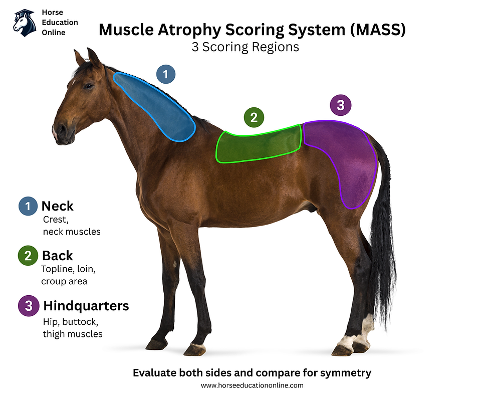
Muscle loss should be documented separately from fat cover. The Equine Body Muscle Score guide explains how to evaluate muscle mass, while Body Condition Scoring assesses body fat. A horse can lose muscle while maintaining or gaining fat, so body weight alone can hide an important change.
Back soreness, weak topline muscles, hindlimb gait changes, and resistance under saddle are not specific to PSSM. The horse may also require investigation for orthopedic pain. Review Back Pain in Horses and Lumbosacral Pain vs. Sacroiliac Pain vs. Hindlimb Lameness for related causes that can resemble or coexist with muscle disease.
Dark Urine Is an Emergency Warning
Severe muscle damage can release myoglobin, a muscle pigment, into the bloodstream and urine. This may turn the urine dark red, brown, or coffee-colored. Myoglobin can contribute to kidney injury, especially when the horse is dehydrated.
Severe pain, dark urine, marked weakness, collapse, or an inability to stand requires immediate veterinary attention. Stop exercise and do not force the horse to walk. Forced movement can increase muscle damage.
If an episode occurs, obtain the horse's temperature, heart rate, respiration rate, gum color, and hydration status if it is safe to do so. The Horse First Aid and Vitals guide and the detailed article on The Horse's Vital Signs explain how to collect useful information before the veterinarian arrives.
Did You Know? Severe tying-up can look like colic. A horse may paw, stretch, sweat, or try to lie down. Firm, painful hindquarter muscles, stiffness, recent exercise, and dark urine raise concern for muscle damage, but an owner cannot safely rule out colic without a veterinary examination.
PSSM vs Tying-Up: What’s the Difference?
PSSM | Tying-Up |
An underlying muscle disorder or group of muscle disorders | A clinical syndrome involving exercise-associated muscle damage |
Can predispose a horse to tying-up episodes | Can have several different underlying causes |
May cause stiffness, weakness, poor performance or muscle loss even without a dramatic episode | Usually refers to an active episode of muscle pain, stiffness and damage |
Requires identifying the specific underlying disorder | Requires determining what caused the episode |
A horse can tie up without having PSSM, and a horse with PSSM may show more subtle signs without experiencing a severe tying-up episode.
What to Do During a Suspected Tying-Up Episode
When a horse becomes stiff, painful, sweaty, or reluctant to move during exercise:
Stop exercise immediately. Do not try to ride through the stiffness.
Do not force the horse to walk. Move only as much as necessary for immediate safety.
Call a veterinarian. Describe the horse's signs, recent work, diet, medications, urine color, and previous episodes.
Keep the horse quiet and comfortable. If nearby, a safe, well-bedded stall or sheltered area may be appropriate. Avoid a long forced walk back to the barn.
Offer fresh water unless the veterinarian advises otherwise. Do not delay the call while trying home treatments.
Check and record vital signs if safe. Note the time the signs began and whether they are improving or worsening.
Do not give medications without veterinary direction. Dehydration and kidney stress can affect the safety of some drugs.
Veterinary treatment depends on severity. It may include pain control, sedation, muscle relaxation, intravenous fluids, correction of acid-base or electrolyte disturbances, and monitoring of kidney function. Horses with myoglobinuria or dehydration may require aggressive fluid support. Learn the practical signs of fluid loss in How to Tell If a Horse Is Dehydrated.
How PSSM Is Diagnosed
PSSM cannot be diagnosed from signs alone. Stiffness, weakness, poor performance, muscle loss, and tying-up occur with many muscle, orthopedic, metabolic, nutritional, and neurologic conditions.
1. History
The veterinarian will review the horse's breed, family history, age at onset, feeding program, exercise schedule, recent rest periods, previous episodes, and response to earlier management changes. The timing of signs in relation to exercise is particularly useful.
Accurate records can reveal patterns that memory misses. The Horse Tracker can be used to log exercise, turnout, feed changes, body condition, muscle loss, veterinary findings, medications, and recurring symptoms.
2. Physical, Lameness, and Neurologic Examination
The examination may include muscle symmetry, muscle firmness and pain, gait at the walk and trot, back and pelvic comfort, neurologic function, hoof pain, and joint-related lameness.
During an acute episode, the veterinarian will also assess heart rate, respiration, hydration, body temperature, and urine color.
A positive PSSM1 result does not prove that every problem is caused by PSSM. Mutation-positive horses can also develop arthritis, hoof pain, back disease, sacroiliac pain, neurologic disease, or another myopathy.
3. Blood Muscle Enzymes
The two enzymes most commonly evaluated are creatine kinase and aspartate aminotransferase.
Creatine kinase, or CK, rises rapidly when muscle cells are damaged. It may increase within hours and fall relatively quickly after active damage stops.
Aspartate aminotransferase, or AST, rises more slowly and may remain increased for several days.
Elevated CK and AST support a diagnosis of muscle injury, but they do not identify its cause. Normal values between episodes do not completely exclude a muscle disorder. In selected cases, a veterinarian may compare enzyme activity before and after controlled exercise. Exercise testing should only be performed under veterinary supervision.
4. Genetic Testing for PSSM1
A validated DNA test can identify the GYS1 mutation using whole blood or mane or tail hairs with intact roots. In an at-risk breed, this is generally the preferred test when PSSM1 is suspected.
A positive result confirms that the horse carries the PSSM1 mutation. It does not predict exactly how severe the signs will be, and it does not prove that PSSM1 is responsible for every performance complaint.
A negative result rules out the known PSSM1 mutation. It does not rule out PSSM2-ER, MFM, recurrent exertional rhabdomyolysis, or another muscle disorder.
5. Muscle Biopsy
A biopsy may be recommended when a horse has recurrent muscle-related signs, blood tests support muscle injury, the PSSM1 test is negative, or PSSM2-ER or MFM is suspected.
The tissue may be evaluated with periodic acid-Schiff, or PAS, staining to highlight glycogen and related carbohydrates. It can then be treated with amylase. Normal glycogen is generally removed, while some abnormal polysaccharide remains visible.
Depending on the suspected disorder, the pathologist may assess:
Total glycogen and its distribution
Amylase-sensitive glycogen
Amylase-resistant polysaccharide
Muscle-fiber degeneration and regeneration
Vacuoles and internal nuclei
Desmin accumulation
Disorganization of myofibrils
Collection, freezing, staining, and interpretation must be performed correctly. Mild glycogen accumulations can be difficult to interpret, so biopsy findings should be considered alongside clinical signs, enzyme activity, breed, and exercise history.

6. Are PSSM2 Genetic Tests Accurate?
Commercial tests marketed with variant names such as P2, P3, P4, or Px should not be treated as validated diagnostic tests for PSSM2 or MFM. Peer-reviewed research found that these variants did not correspond reliably with biopsy-confirmed disease.
These panels should not be used alone to:
Diagnose PSSM2 or MFM
Explain poor performance
Predict prognosis
Exclude a horse from breeding
Make prepurchase decisions
At present, PSSM1 is the form for which a scientifically validated commercial genetic test is available. A horse with symptoms and a negative PSSM1 test still needs a proper veterinary investigation.
Did You Know? A genetic result and a clinical diagnosis answer different questions. The PSSM1 test identifies the known GYS1 mutation. Blood enzymes show whether muscle damage has occurred. A biopsy can describe changes within muscle tissue. No single result replaces the complete clinical assessment.
Conditions That Can Resemble PSSM
Important differential diagnoses include:
Sporadic or recurrent exertional rhabdomyolysis
Myofibrillar myopathy
Malignant hyperthermia
Myosin heavy-chain myopathy
Hyperkalemic periodic paralysis
Electrolyte abnormalities
Vitamin E or selenium deficiency
Infectious or inflammatory muscle disease
Toxic muscle damage
Overexertion or inadequate conditioning
Orthopedic lameness
Back or sacroiliac pain
Neurologic disease
The management and prognosis of these conditions differ substantially. Labeling every stiff or poorly performing horse as PSSM can delay the diagnosis of a treatable orthopedic, neurologic, nutritional, or systemic problem.
Long-Term Management of PSSM1
PSSM1 is lifelong. The mutation cannot be removed, but many horses can remain comfortable and return to useful work when their program is consistent.
Build the Diet Around Forage and Caloric Need
The feeding plan should begin with the horse's body weight, body condition, workload, forage analysis, and total calorie requirement. Many PSSM1 horses are easy keepers. Adding fat without first calculating energy intake can create obesity, which adds a separate health and performance problem.
General management commonly includes:
Good-quality forage as the foundation of the diet
Restriction of high-starch and high-sugar feeds
A low-nonstructural-carbohydrate ration balancer when additional calories are not required
A carefully selected low-starch, higher-fat feed when more energy is needed
Adequate high-quality protein, vitamins, minerals, salt, and water
Regular reassessment of weight and body condition
The precise target for starch, sugar, fat, and forage depends on how the ration is calculated and on the individual horse. Work with a veterinarian or qualified equine nutritionist rather than choosing a feed from the front label alone. The Basics of Equine Nutrition provides a useful foundation for understanding forage, concentrates, protein, energy, vitamins, and minerals.
Use the Horse Weight and Body-Condition Estimator to follow trends, but confirm important feeding decisions with hands-on body condition scoring and professional advice.
Exercise Consistency Matters
Diet alone is often insufficient. Regular exercise improves the muscle's ability to use stored energy, and daily turnout reduces the long periods of inactivity that can precede an episode.
A management program may include:
As much safe daily turnout as practical
A gradual return to work after an episode
A long, controlled warm-up
Low-intensity work introduced before speed, collection, hills, or demanding strength exercises
Slow increases in duration and intensity
Avoidance of several days of complete rest followed by a full workout
Reduction of feed calories when the workload decreases
The veterinarian should determine when exercise can safely resume after an acute episode. The schedule may depend on pain, hydration, kidney function, CK and AST results, and the severity of muscle damage.
Track the Whole Horse
Record more than tying-up episodes. Useful observations include body weight, body condition, muscle score, appetite, urine color, turnout time, type and duration of exercise, warm-up, weather, transport, rest days, and changes in feed. Trends may identify a trigger or show that another disease process is emerging.
Sweating horses need appropriate access to water, salt, and electrolytes, but electrolyte products do not treat the underlying glycogen-storage disorder. The Equine Salt and Electrolyte Calculator can help estimate routine needs based on work and climate. Use veterinary guidance during illness, dehydration, or active rhabdomyolysis.
Management of PSSM2-ER and Myofibrillar Myopathy
Some Quarter Horse-related horses with biopsy-confirmed PSSM2-ER improve with the same broad principles used for PSSM1, including consistent exercise, turnout, gradual conditioning, and an individualized low-starch feeding program. The response is less predictable because the cause and full clinical range remain incompletely understood.
Warmbloods and Arabians with MFM or another GYS1-negative myopathy require individualized management. Some improve with controlled exercise, avoidance of prolonged rest, careful conditioning, dietary adjustment, and treatment of concurrent orthopedic pain. Others continue to show muscle loss, gait abnormalities, or poor performance.
The feeding strategy used for PSSM1 should not automatically be applied indefinitely to every Warmblood or Arabian labeled PSSM2. If the horse fails to improve, the diagnosis and ration should be reassessed with the veterinarian and nutritionist. The term PSSM2 does not guarantee a single disease mechanism or a single correct diet.
Did You Know? Improvement after a diet change does not confirm a diagnosis. A horse may improve because calories, protein, forage quality, turnout, training consistency, gastric comfort, or another part of management changed at the same time.
PSSM and Breeding Decisions
Because PSSM1 is autosomal dominant, a horse with one copy has approximately a 50 percent chance of transmitting the mutation to each foal. A horse with two copies will transmit it to every foal. Each breeding is a new probability event.
Validated PSSM1 results can therefore provide meaningful information before breeding.
Owners should discuss the horse's genotype, clinical history, family, intended offspring, and breed recommendations with a veterinarian and breed organization.
The situation is different for PSSM2-ER and MFM. No causative commercial variant has been validated for these conditions. Unvalidated P2, P3, P4, or Px panels should not be used as the sole basis for removing a horse from a breeding program or predicting an offspring's risk. When a horse has biopsy-confirmed disease or a strong family pattern, seek advice from a veterinarian with expertise in equine neuromuscular disease and genetics.
Prognosis
The prognosis for life is generally favorable for many horses with PSSM1. The prognosis for athletic use depends on the severity of previous episodes, consistency of exercise, access to turnout, dietary management, genotype, breed, intended workload, and the presence of other disease.
Many horses improve substantially when both feeding and exercise recommendations are followed. A horse with mild episodes that responds well to management may return to useful or competitive work. Clinical signs may recur when the program is interrupted, especially after rest followed by sudden exercise.
The prognosis becomes more guarded with:
Repeated severe rhabdomyolysis
Persistent elevation of muscle enzymes
Progressive muscle atrophy
Marked weakness
Recumbency
Kidney injury
Poor response to careful management
Another muscle-disease mutation
Inability to provide consistent exercise or turnout
Severe and occasionally fatal disease has been documented in Belgian and Percheron Draft Horses, although this is not the expected outcome for every affected draft horse.
Prognosis for PSSM2-ER is harder to predict because the causative mutation and full spectrum of disease are unknown. Outcomes in horses historically diagnosed with PSSM2 are also variable because the category may include MFM and other incompletely defined conditions.
What the Evidence Supports, and What Remains Uncertain
Strong evidence supports that PSSM1 is caused by a gain-of-function GYS1 mutation, is inherited as an autosomal dominant trait, can be identified through validated DNA testing, and causes excessive glycogen and abnormal polysaccharide accumulation in skeletal muscle. Diet and consistent exercise affect clinical expression.
Research also shows that commercial P2, P3, and P4 variants do not reliably correspond with biopsy-confirmed PSSM2 or MFM.
Evidence is developing for PSSM2-ER as a genuine GYS1-negative glycogen-storage disorder in Quarter Horse-related breeds. However, its specific genetic cause remains unknown. The cause of MFM in Warmbloods and Arabians, the best treatment for every GYS1-negative horse, and the clinical significance of mild biopsy changes also remain incompletely understood.
Clear communication is important. When a horse is called PSSM-positive, ask whether that means a validated PSSM1 test, a biopsy diagnosis, or a commercial variant-panel result. Those are not interchangeable findings.
Frequently Asked Questions About PSSM in Horses
What is PSSM in horses?
PSSM, or polysaccharide storage myopathy, is a group of equine muscle disorders involving abnormal storage of glycogen or related polysaccharides within skeletal muscle.
Affected horses may show stiffness, muscle pain, exercise intolerance, weakness, poor performance, muscle loss or episodes of tying-up. The Interactive Horse Muscles Tool can help owners understand the major muscle groups involved.
What is the difference between PSSM1 and PSSM2?
PSSM1 is a defined inherited disorder caused by a mutation in the GYS1 gene and can be identified with a validated DNA test.
The term PSSM2 has historically been used for several GYS1-negative muscle disorders. Current evidence indicates that this category is not one single disease and may include PSSM2-ER in Quarter Horse-related breeds and myofibrillar myopathy in some Warmbloods and Arabians.
What are the first signs of PSSM in a horse?
Early signs may include stiffness shortly after exercise begins, a shortened stride, reluctance to move forward, firm or painful hindquarter muscles, unexplained sweating, muscle tremors or declining performance.
Some horses develop more subtle exercise intolerance or muscle loss. The Equine Body Muscle Score guide can help document muscle changes separately from body fat.
Is PSSM the same as tying-up?
No. Tying-up, or exertional rhabdomyolysis, is a syndrome involving exercise-associated muscle damage.
PSSM can make a horse more susceptible to tying-up, but other conditions can cause similar episodes. A horse with stiffness or muscle pain should not automatically be assumed to have PSSM.
How is PSSM diagnosed?
PSSM cannot be diagnosed from symptoms alone.
Diagnosis may involve the horse's history, physical examination, lameness and neurologic assessment, blood testing for CK and AST muscle enzymes, validated genetic testing for PSSM1 and muscle biopsy in selected cases.
A positive PSSM1 test confirms that the horse carries the GYS1 mutation, but it does not prove that every performance or gait problem is caused by PSSM.
Is there a reliable genetic test for PSSM2?
No scientifically validated commercial DNA test currently diagnoses PSSM2-ER or myofibrillar myopathy.
Commercial tests marketed as P2, P3, P4 or Px should not be used alone to diagnose PSSM2, explain poor performance, predict prognosis or make breeding decisions.
PSSM1 is currently the form with a validated commercial genetic test.
What should you feed a horse with PSSM1?
Management commonly begins with good-quality forage and restriction of high-starch and high-sugar feeds.
Some easy keepers may need only appropriate forage and a low-NSC ration balancer, while horses requiring additional calories may benefit from a carefully selected low-starch, higher-fat feed. The ration should be based on body weight, workload, forage analysis and professional guidance.
The Basics of Equine Nutrition provides useful background on balancing forage, energy, protein, vitamins and minerals.
What should you do if a horse with suspected PSSM starts tying up?
Stop exercise immediately and contact a veterinarian. Do not force the horse to continue walking because additional movement may increase muscle damage.
Keep the horse quiet and comfortable, offer fresh water unless advised otherwise and record vital signs if it is safe to do so. The guide to horse vital signs can help owners collect useful information before the veterinarian arrives.
Dark red, brown or coffee-colored urine, severe pain, marked weakness, collapse or inability to stand requires immediate veterinary attention.
Can a horse with PSSM still be ridden?
Many horses with well-managed PSSM1 can return to regular or competitive work.
Consistent exercise, turnout, gradual conditioning and controlled warm-ups are important parts of management. Several days of complete rest followed by a full workout can increase the risk of an episode.
Horse Tracker can be used to record exercise, turnout, feed changes, rest days, body condition, muscle score, laboratory results and recurring symptoms.
Key Takeaways
PSSM affects skeletal muscle and may cause exercise-associated muscle pain and damage.
PSSM1 is caused by a validated, dominantly inherited mutation in the glycogen synthase 1 gene, GYS1.
A DNA test using blood or mane or tail hairs with intact roots can identify PSSM1.
A negative PSSM1 test does not rule out another muscle disorder.
The label PSSM2 has historically included several conditions that may not share the same cause.
Commercial P2, P3, P4, and Px variant panels have not been shown to diagnose biopsy-confirmed PSSM2 or myofibrillar myopathy reliably.
Diet, turnout, and consistent exercise can strongly influence the expression of PSSM1, but they do not cause or remove the genetic mutation.
Severe pain, dark urine, marked weakness, collapse, or an inability to stand requires immediate veterinary attention.
Final Thoughts
PSSM is best understood as a group of muscle disorders rather than a single diagnosis. PSSM1 has a confirmed genetic cause and a validated DNA test. PSSM2-ER is a GYS1-negative glycogen-storage disorder recognized in Quarter Horse-related breeds, but its genetic cause has not been identified. Some Warmbloods and Arabians formerly labeled PSSM2 may instead have myofibrillar myopathy involving structural muscle proteins.
Accurate diagnosis protects the horse from oversimplified treatment. A genetic test, blood enzymes, muscle biopsy, clinical examination, feeding history, and exercise pattern each provide different information. Many horses with PSSM1 can have useful, active lives when diet and exercise are managed consistently, but any new pain, gait change, weakness, or performance decline deserves a fresh veterinary assessment.
Browse additional practical guides in the Horse Health Conditions library, and use the Horse Tracker to keep diet, exercise, symptoms, laboratory results, and management changes together.
This article is for educational purposes and is not a substitute for diagnosis or treatment by a veterinarian.
Selected Peer-Reviewed References
Firshman AM, Valberg SJ. Polysaccharide Storage Myopathy. Veterinary Clinics of North America: Equine Practice. 2025;41(1):125-137. https://doi.org/10.1016/j.cveq.2024.11.004
McCue ME, Valberg SJ, Miller MB, et al. Glycogen synthase GYS1 mutation causes a novel skeletal muscle glycogenosis. Genomics. 2008;91(5):458-466. https://doi.org/10.1016/j.ygeno.2008.01.011
McCue ME, Valberg SJ, Lucio M, Mickelson JR. Glycogen synthase 1 mutation in diverse breeds with polysaccharide storage myopathy. Journal of Veterinary Internal Medicine. 2008;22(5):1228-1233. https://doi.org/10.1111/j.1939-1676.2008.0167.x
Valberg SJ, Williams ZJ, Finno CJ, et al. Type 2 polysaccharide storage myopathy in Quarter Horses is a novel glycogen storage disease causing exertional rhabdomyolysis. Equine Veterinary Journal. 2023;55(4):618-631. https://doi.org/10.1111/evj.13876
Valberg SJ, Finno CJ, Henry ML, et al. Commercial genetic testing for type 2 polysaccharide storage myopathy and myofibrillar myopathy does not correspond to a histopathological diagnosis. Equine Veterinary Journal. 2021;53(4):690-700. https://doi.org/10.1111/evj.13345
Williams ZJ, Valberg SJ, et al. Clinical and histopathological features of myofibrillar myopathy in Warmblood horses. Equine Veterinary Journal. 2017;49(6):728-735.
Firshman AM, Valberg SJ, Bender JB, et al. Epidemiologic characteristics and management of polysaccharide storage myopathy in Quarter Horses. American Journal of Veterinary Research. 2003;64(10):1319-1327.
Hunt LM, Valberg SJ, Steffenhagen K, McCue ME. An epidemiological study of myopathies in Warmblood horses. Equine Veterinary Journal. 2008;40(2):171-177.


Comments