
Chronic Progressive Lymphedema in Draft Horses


Signs, Causes, Treatment, and Lifelong Management
Chronic progressive lymphedema, commonly shortened to CPL, is a lifelong disease affecting the lower limbs of many heavy draft horses and other heavily feathered breeds. It causes persistent swelling, thickened skin, scaling, folds, nodules, sores, and repeated infections. Advanced disease may interfere with movement, hoof care, and quality of life.
CPL is easy to overlook in its early stages. Long feathering can conceal swelling and skin folds, and the first crusts may be dismissed as ordinary scratches or mud fever. By the time the changes are clearly visible from a distance, permanent fibrosis may already have developed.
There is currently no cure for CPL. Early recognition and consistent care can still make a meaningful difference. Management is aimed at slowing progression, reducing swelling, controlling mites and secondary infections, protecting the skin, and keeping the horse comfortable and mobile for as long as possible.
Owners who are uncertain whether a lower-leg problem resembles mud fever, rain rot, or another common skin condition can begin with the Horse Skin Conditions Decision Helper. A veterinarian is still needed to diagnose CPL and identify its complications.
What Is Chronic Progressive Lymphedema?
CPL is a chronic disorder involving the skin, connective tissue, and lymphatic system of the distal limbs. The distal limb is the part of the leg farthest from the body, generally extending from the cannon region downward. All four legs may be affected, although disease severity is not always equal from one leg to another.
The name describes three features:
Chronic: It persists over a long period and requires lifelong attention.
Progressive: The tissue changes usually become more severe with age.
Lymphedema: Lymphatic fluid accumulates because it is not being cleared normally.
CPL has also appeared in older literature as chronic pastern dermatitis or chronic proliferative pastern dermatitis. Those terms describe the visible skin lesions, but changes in the lymphatic system and deeper connective tissues are central to the condition.
The role of the lymphatic system
The lymphatic system is a network of small vessels that collects excess fluid, proteins, inflammatory material, and cellular waste from tissues. It has no heart-like central pump.

Limb movement, muscle contraction, changing tissue pressure, and the elasticity of surrounding structures help propel lymphatic circulation.
When drainage is impaired, protein-rich fluid remains in the tissues and sustains inflammation. Over time, the body produces increasing amounts of dense connective tissue.
This process is called fibrosis. Flexible tissue is gradually replaced or infiltrated by stiff, scar-like connective tissue. Early edema may feel soft and indent when pressed. Advanced CPL often feels firm because fibrotic tissue contributes to the enlargement. Exercise may reduce the fluid component, but it cannot reverse established fibrosis.
Did You Know? A hard, enlarged lower leg is not necessarily holding only fluid. In advanced CPL, permanent fibrous tissue contributes substantially to the size and firmness of the limb. This is one reason late-stage swelling does not disappear after turnout or exercise.
Which Horses Are Most Commonly Affected?
CPL is primarily recognized in heavy, feathered breeds. Reported susceptible breeds include:
Belgian Drafts
Shires
Gypsy Cobs and Gypsy Vanners
Percherons
Ardennes
Bretons and Boulonnais
Rhenish German, Schleswig, South German, Black Forest, and other German draft breeds
Crossbred horses descended from susceptible breeds
The Belgian Draft has been studied most extensively. Some examined populations have shown extremely high prevalence, but those figures should not be applied automatically to every farm, bloodline, country, or breed. Prevalence changes with the age of the horses, the population sampled, and how carefully the legs are clipped, palpated, and scored.
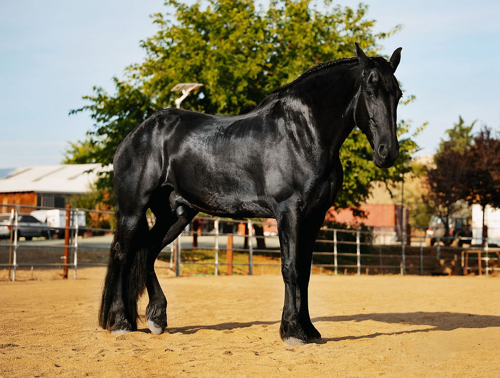
The condition is also documented in Friesians. Owners of this breed can learn more about its broader history, characteristics, and health considerations in the article: The Friesian Horse.
Heavy feathering has not been proven to cause CPL by itself, although it makes detection and treatment harder. Signs usually become more obvious with age as damage accumulates. A young horse may look normal yet develop clear disease later. CPL can affect both sexes, although males have been more severely affected in some studied populations.
Did You Know? CPL may begin before it can be seen. Careful palpation can find early thickening and small folds beneath dense feathering when the leg still looks normal from a few feet away.
What Causes CPL?
The initiating cause has not been identified. CPL is considered multifactorial. Genetics, lymphatic dysfunction, abnormal elastin, inflammation, and fibrosis are connected, but researchers have not established which change comes first.
1. Inherited susceptibility
CPL repeatedly occurs in particular breeds and bloodlines. Researchers have identified associated chromosome regions, but no single mutation explains every case. There is no definitive commercial DNA test that can declare a horse free of CPL or predict its severity.
A 2024 study of Rhenish German Draft horses found substantial heritability for CPL severity. One model estimated approximately 0.60 across all ages, with results varying by model and age restrictions. This means genetic differences explained a substantial share of variation within that studied population. It does not mean that 60 percent of an individual horse's disease was caused by its genes.
Care still matters. Mite control, skin care, movement, wound prevention, and prompt infection treatment can limit secondary damage, but they cannot remove inherited susceptibility.
2. Abnormal elastin and connective-tissue support
Elastin is a flexible protein in skin, blood vessels, and tissues around lymphatic structures. Research has found differences in the amount, structure, or metabolism of dermal elastin in affected draft horses.
Elastic fibers help keep early lymphatic channels open and support tissue-fluid uptake. An abnormal elastin network may reduce lymph clearance, although researchers do not know whether elastin disruption begins CPL, results from inflammation, or works in both directions.
3. Reduced lymphatic clearance
Lymphatic imaging has demonstrated reduced lymph clearance in affected draft-horse limbs. Retained fluid and proteins encourage inflammation, tissue thickening, and connective-tissue production.
The lower limbs contain little muscle below the knee and hock and depend heavily on movement to assist fluid return. Prolonged standing can make swelling more noticeable when lymphatic vessels are compromised.
4. Chronic inflammation and fibrosis
Continued inflammation stimulates excessive connective-tissue production. The resulting fibrosis distorts or compresses lymphatic pathways, making drainage less effective.
The disease can therefore become self-perpetuating:
Reduced lymphatic drainage → protein-rich swelling → inflammation → fibrosis → further loss of lymphatic function
Cracks, ulcers, mites, infection, moisture, and rubbing add further inflammation. Treating them is important, but the underlying cycle remains.
Did You Know? Researchers agree that lymphatic dysfunction, inflammation, fibrosis, and abnormal elastin are involved in CPL. What remains uncertain is their exact order. Lymphedema may drive inflammation, while inflammation and fibrosis can also make lymphatic drainage worse.
Factors That Aggravate CPL
Several problems accelerate skin damage or make CPL harder to control. Finding and treating them is central to long-term management.
Chorioptes mites
Chorioptes bovis, commonly called the feather or leg mite, lives on the skin surface and feeds primarily on debris. Infested horses may stamp, rub, bite their legs, repeatedly lift their feet, or become restless. Hair loss, scale, crusts, thickening, and self-inflicted wounds may follow.

In a 2025 Belgian study of 156 Belgian Draft horses and 142 Belgian Warmbloods, researchers found Chorioptes mites in 92.31 percent of the Drafts and CPL in 80.77 percent. Neither was found in the Warmblood group. Mites and CPL were strongly associated within the studied Draft population.
The association does not prove that mites are the sole cause. CPL has a broader inherited and tissue-level basis, while mites amplify disease through inflammation, itching, stamping, biting, and rubbing.
Mites may be hidden within heavy crusts and thick folds. A negative superficial skin scraping does not always rule them out because the sample may miss an unevenly distributed population.
Did You Know? In the 2025 Belgian study, mites were found in Draft foals as young as six days old. CPL and mite prevalence rose sharply with age in that population, showing why prevention and careful examination should begin early.
Secondary bacterial and fungal infections
Deep folds trap moisture and debris. Cracked or ulcerated skin allows bacteria and fungi to enter, increasing swelling, odor, discharge, heat, pain, and tissue damage. Deep infection may produce cellulitis or lymphangitis.
A veterinarian may need cytology, bacterial culture and sensitivity, or fungal testing when lesions are deep, recurrent, spreading, or unresponsive.
Feathering, moisture, and trapped debris
Dense hair can conceal CPL, retain moisture and debris, prevent topical treatment from reaching the skin, and make folds difficult to dry. Mud and standing water worsen maceration, the softening and breakdown of skin caused by prolonged moisture.
CPL should not be confused with ordinary mud fever or pastern dermatitis. Both can produce crusts, sores, and swelling around the pastern, and they may occur together. CPL is distinguished by its progressive lymphatic dysfunction, characteristic firm thickening, folds, and nodules in a susceptible horse.
Reduced movement
Walking creates pressure changes through the limb and hoof that help return blood and lymph. Confinement, lameness, obesity, or pain that discourages movement can increase dependent swelling.
Exercise is supportive and cannot remove fibrosis. It may be unsafe during severe pain, acute cellulitis, or deep wounds.
Trauma and friction
Thick folds and nodules are vulnerable to rubbing, tearing, and pressure. Ill-fitting boots, abrasive footing, stamping, clippers, or a tight bandage may open the skin. Swollen, stiff tissue often heals slowly.
Clinical Signs of CPL
CPL usually develops gradually. The earliest changes may be found only by parting the hair and feeling the entire circumference of each lower limb. Owners should compare opposite legs while remembering that both sides may be affected.
Early signs
Mild swelling around the pastern, fetlock, or lower cannon region
Skin that feels thicker or less mobile than expected
A small transverse fold behind the pastern
Scaling or persistent dandruff beneath the feathers
Recurrent crusting that does not respond as expected to routine pastern care
Mild itching, stamping, rubbing, or foot lifting
Swelling that improves somewhat with movement but never fully resolves
Subtle hair loss or broken feathering
At this stage, the horse may remain sound and comfortable. The absence of lameness does not mean the tissue is normal.
Progressive signs
Enlarging folds around the back and sides of the pastern
Firm or hard thickening of the skin and subcutaneous tissues
Raised, wart-like nodules
Excessive scale and heavy crust formation
Cracks, erosions, and ulcers hidden within folds
Moist or foul-smelling discharge
Repeated mite infestations
Recurrent bacterial or fungal dermatitis
Increasing circumference of the lower limb
Reduced flexibility when the fetlock is flexed
Advanced signs
Large, hard folds extending upward along the cannon region
Numerous nodules or masses on several surfaces of the limb
Deep, chronically infected wounds
Severe fibrosis with little normal skin mobility
Pain when the limb is touched, flexed, or lifted
Difficulty lifting the feet for trimming or shoeing
Altered gait, lameness, or reluctance to move
Poor hoof quality or distortion associated with reduced movement and uneven loading
Recurrent episodes of cellulitis or lymphangitis
Declining body condition, activity, and quality of life in severely affected horses
Owners can use the Comprehensive Guide to Equine Lameness to become more observant of stance and movement, but a painful or rapidly swollen limb should be examined by a veterinarian rather than subjected to repeated owner testing.

How CPL Is Diagnosed
No single blood, skin, or genetic test confirms every CPL case. Diagnosis begins with breed, age, family history, appearance, and hands-on examination of all four lower limbs.
A thorough evaluation may include:
Parting or clipping the feathers so the skin can be seen
Palpating the complete circumference of each lower limb
Assessing tissue thickness, mobility, temperature, pain, and firmness
Identifying folds, nodules, scale, crusts, ulcers, and discharge
Comparing the pattern and severity among all four legs
Watching the horse move and evaluating comfort during limb flexion
Looking for mites and secondary infection
Reviewing the horse's history of recurrent pastern dermatitis or unexplained swelling
The Interactive Horse Skeleton can help owners and students review the structures of the lower limb and communicate lesion locations more accurately to a veterinarian or farrier.
Additional diagnostic tests
Testing is chosen according to the lesions and differential diagnoses:
Superficial skin scrapings: Used to look for mites, although several samples or repeat testing may be necessary.
Cytology: Examines cells, bacteria, yeast, and inflammation collected from the skin.
Bacterial culture and sensitivity: Helps select an antibiotic when infection is deep, recurrent, severe, or previously treated.
Fungal testing: Used when fungal dermatitis is suspected.
Skin biopsy: May help in unusual cases or when immune-mediated, neoplastic, or other disorders are possible.
Ultrasound: Can demonstrate thickening of skin and subcutaneous tissues or investigate another cause of swelling.
Lymphatic imaging: Used mainly in research or specialist settings to assess lymph clearance.
Bloodwork: Does not diagnose CPL directly, but may help evaluate systemic illness, infection, protein loss, or another reason for limb swelling.
Conditions that can resemble CPL
A veterinarian may need to distinguish CPL from:
Pastern dermatitis caused primarily by moisture, friction, photosensitization, or contact irritation
Chorioptic mange without established CPL
Acute cellulitis or lymphangitis
Dermatophilosis or fungal infection
Vasculitis
Pemphigus or another immune-mediated skin disease
Local trauma or an infected wound
Limb swelling related to heart, liver, kidney, vascular, or protein-loss disorders
An acutely hot, painful, rapidly swollen leg with fever is not typical of stable CPL. It may indicate cellulitis, lymphangitis, or another urgent complication.
Treatment and Long-Term Management
Can CPL be cured?
There is currently no cure for chronic progressive lymphedema. Treatment cannot reliably restore severely damaged lymphatic vessels or convert mature fibrotic tissue back into normal flexible skin. The practical goals are to:
Slow further progression
Reduce the fluid component of swelling
Maintain skin flexibility and integrity
Control mites
Identify and treat infection
Protect wounds from contamination and trauma
Preserve comfortable movement
Maintain regular hoof care
Reduce pain and support quality of life
The plan must be individualized. A routine that is suitable for dry, intact skin may be harmful when the same horse has an active infection, painful ulcer, or acute cellulitis.
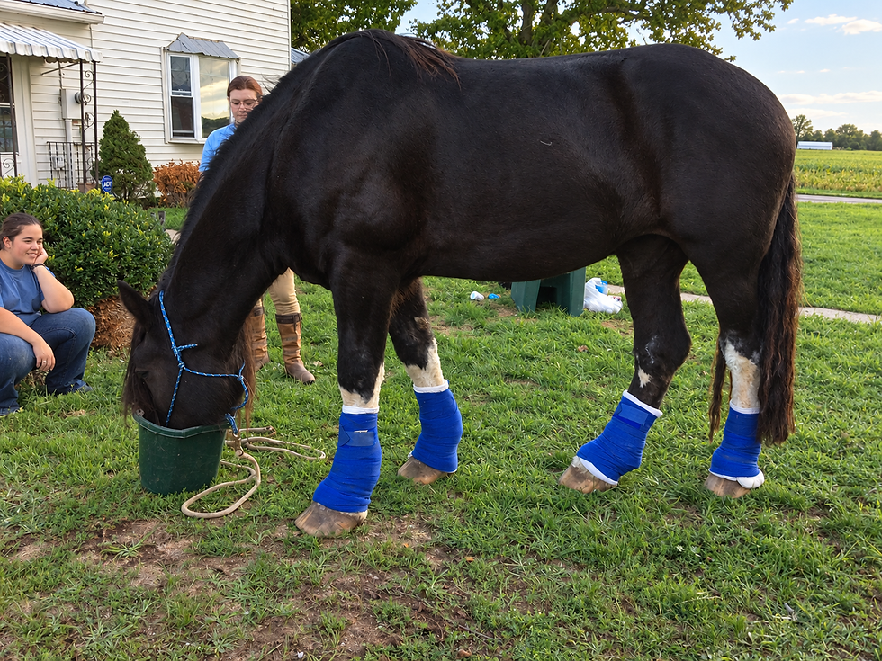
1. Expose the skin for examination
Long feathers often need to be clipped or shortened so the skin can be examined, cleaned, dried, and medicated. Folded, tender skin is easy to cut, so use clean equipment, keep blades cool, and never run a blade blindly beneath a mat or across a nodule. Professional handling or veterinary pain control may be needed. Access to active disease takes priority over preserving feather for presentation.
2. Clean gently and only as needed
Remove loose scale, discharge, and debris without creating new wounds. A veterinarian may recommend an antimicrobial or keratolytic cleanser. Keratolytics soften excessive keratin and dead skin and may contain ingredients such as urea, sulfur, salicylic acid, or benzoyl peroxide. They are not interchangeable and may irritate raw areas.
Do not scrub aggressively or pull off attached crusts. Soften debris as directed and remove only what releases easily. Repeated washing is counterproductive when it keeps the leg wet.
3. Dry every fold completely
Moisture beneath hair, crusts, or nodules encourages maceration and microbial growth. After washing, use clean towels and, if tolerated, a cool or mildly warm air source. Avoid excessive heat.
Pay close attention to:
The back of the pastern
The heel bulbs and coronary band
Folds around the fetlock
The undersides of nodules
The back of the knee
The front of the hock
Do not share towels or grooming tools between affected horses.
4. Protect flexibility without sealing in infection
Dry, rigid skin splits more easily during movement, so a veterinarian may recommend an emollient for clean, dry, noninfected areas. Thick grease can trap moisture, dirt, mites, and discharge or conceal deterioration. Do not cover wet skin or untreated infection unless directed.
5. Control Chorioptes mites
Mite treatment should be designed by a veterinarian. Livestock products may be unsafe for horses, and many equine Chorioptes treatments are off label. Scale, folds, and feathers can block topical medication, while other horses, equipment, or the environment may cause reinfestation.
A controlled 2023 study evaluated a high-dose topical moxidectin pour-on protocol after clipping in 19 CPL-affected Belgian Draft horses. Detectable live mites were eliminated after treatment, itching and mite-associated lesions improved, and some CPL skin lesions improved. Established folds did not significantly shrink, and some horses were later reinfested.
The study supports mite control but is not a dosing guide. Its formulation, dose, route, clipping, timing, and monitoring require veterinary interpretation.
A complete mite-control plan may involve:
Clipping enough hair for medication to reach the skin
Softening or removing obstructive crusts without injuring the horse
Treating all affected areas at the prescribed interval
Examining or treating in-contact horses as directed
Cleaning stalls, grooming areas, and trailers
Laundering or disinfecting brushes, towels, wraps, and clippers
Avoiding shared leg-care equipment
Repeating skin scrapings when appropriate
Medication warning: Amitraz should not be used on horses because serious toxic effects have been reported. Never apply a livestock acaricide, pesticide concentrate, or improvised chemical treatment without veterinary direction.
6. Treat confirmed infection
Antibiotics treat secondary bacterial infection, not CPL. Fungal problems and mites require different medication. Culture-directed therapy is especially important when infection is deep, widespread, recurrent, previously treated, or accompanied by marked pain, discharge, fever, or swelling. Severe cases may need systemic medication and professional wound care.
7. Encourage regular, controlled movement
Daily turnout, hand-walking, light riding, driving, or controlled ground exercise can assist lymphatic return and maintain flexibility. A horse with acute cellulitis, marked lameness, deep ulcers, or severe pain needs veterinary guidance. The aim is comfortable movement, not forced exertion.
Owners should watch for shorter steps, reluctance to turn, difficulty crossing the limbs, or resentment when a foot is lifted. The Equine Lameness Study Kit and Lameness Flashcards provide structured learning for owners, farriers, and students who want a deeper understanding of gait assessment and limb disease.
8. Consider professional lymphatic therapy and compression
Manual lymphatic drainage is a specialized, gentle technique intended to direct fluid toward functioning pathways. It is not deep massage, which may injure inflamed, infected, painful, or ulcerated tissue.
A pilot study of five affected draft horses combined lymphatic drainage, specialized compression, exercise, and skin or infection management. After two weeks, mean lower-limb volume decreased by about 11.25 percent, with reported improvements in skin and movement. Mites, infection, and unrelated lameness required treatment adjustments.
Five horses are not enough to establish a standard protocol, and temporary improvement is not a cure. Treatment requires proper training and veterinary coordination.
Specialized compression uses specific materials, graduated pressure, padding, and monitoring. It is not an ordinary standing wrap. Poor application can create pressure over a nodule, injure skin, trap moisture, conceal infection, or impair circulation.
9. Manage wounds promptly
Open sores should be protected from contamination, flies, friction, and additional trauma. The wound plan may include gentle cleansing, veterinarian-selected dressings, controlled bandaging, pain relief, culture, and treatment of infection.
Monitor for:
Increasing heat or swelling
Worsening pain
Foul odor or heavier discharge
Dead, gray, or black tissue
Bleeding that does not stop normally
Fly eggs or maggots
Extension into a deeper fold
Fever, depression, or loss of appetite
Persistent or unusual wounds may require biopsy, advanced dressings, surgical assessment, or referral.
10. Reserve surgery for selected lesions
Surgery may be considered when a large nodule repeatedly tears or bleeds, prevents movement, traps infection, causes pain, or blocks farrier care. It is not a routine cure. Lower-limb healing can be difficult, exuberant granulation tissue may develop, and lesions may return. The decision must account for expected aftercare.
11. Maintain regular hoof care
CPL can alter stance, movement, and weight bearing. Painful folds make limb lifting difficult, while reduced movement can contribute to hoof distortion. Regular trimming or shoeing supports balanced loading and allows repeated inspection.
Tell the farrier about pain, wounds, infection, reduced flexion, or medication affecting handling. An advanced horse may need shorter appointments, rest breaks, altered limb positions, or veterinary pain management.
Owners can review the principles of hoof balance and use the Hoof Anatomy Flashcards to better understand the structures being managed.
Building a Practical CPL Care Routine
Write the care schedule with the veterinarian and adjust it whenever the skin changes.
Daily or frequent checks
Watch the horse walk and turn before handling the legs.
Note stamping, rubbing, biting, or repeated foot lifting.
Feel for new heat, pain, soft swelling, hard thickening, or moisture.
Part the hair and inspect established folds and nodules.
Check open areas for odor, discharge, flies, and tissue color.
Confirm that treated skin and bandages remain clean and dry.
Provide comfortable movement when appropriate.
Each week, photograph the legs from consistent angles, compare measurements if the veterinarian recommends them, check equipment for rubbing or moisture, and inspect high-traffic footing for mud.
Records that matter
Record lesion locations, veterinary findings, medication details, mite treatments, cultures, photographs, lameness, and farrier accommodations. The Horse Tracker can keep medical and hoof-care records together.
Did You Know? Familiarity can make slow progression difficult to see. Photographs taken in the same light and position often reveal changes in fold size, skin texture, and limb circumference that are missed during daily handling.
Important Precautions
Do not assume every crust is ordinary scratches
Persistent pastern dermatitis in a susceptible breed deserves examination for folds, edema, mites, and fibrosis.
Do not use antibiotics without a diagnosis
Antibiotics cannot restore lymphatic function or remove fibrosis. Poorly targeted use contributes to resistance and may miss the actual complication.
Avoid harsh chemicals and forceful scrubbing
Concentrated bleach, strong acids, undiluted disinfectants, caustic hoof products, and livestock pesticides can burn the skin. Forceful scrubbing and crust removal create fresh injuries.
Do not leave folds wet
If the leg must be washed, it must also be dried thoroughly. Trapped water beneath a greasy product or wrap can worsen maceration.
Do not apply tight ordinary wraps
Irregular folds and nodules create pressure points. Compression therapy requires a trained professional and is not equivalent to pulling a standing wrap more tightly.
Prevent cross-contamination
Do not share brushes, towels, wraps, or clippers when mites or infection are suspected. Handle unaffected horses first when practical.
Monitor weight and mobility
Excess body weight increases the effort required for movement and may make hoof handling more difficult. Weight loss should be gradual and based on a balanced ration. The Horse Weight and Body-Condition Estimator and guide to Body Condition Scoring can help owners track trends objectively.
When Veterinary Attention Is Urgent
Contact a veterinarian promptly if a horse with known or suspected CPL develops:
Fever
Sudden or rapidly increasing swelling
A hot, very painful limb
Rapidly spreading redness
Severe or non-weight-bearing lameness
Foul-smelling or heavy discharge
Deep ulcers or exposed deeper tissue
Gray or black tissue
Uncontrolled bleeding from a nodule
Fly larvae or maggots in a wound
Depression, loss of appetite, or unusual quietness
Inability to walk comfortably
Swelling that rapidly extends above the previously affected area
These signs may indicate cellulitis, acute lymphangitis, severe infection, tissue necrosis, or another emergency occurring in addition to CPL. Learn the horse's normal baseline before a crisis by reviewing The Horse's Vital Signs and How to Tell if Your Horse Is Sick.
Prognosis
Prognosis depends on disease stage, control of mites and infection, mobility, and the owner's ability to continue care.
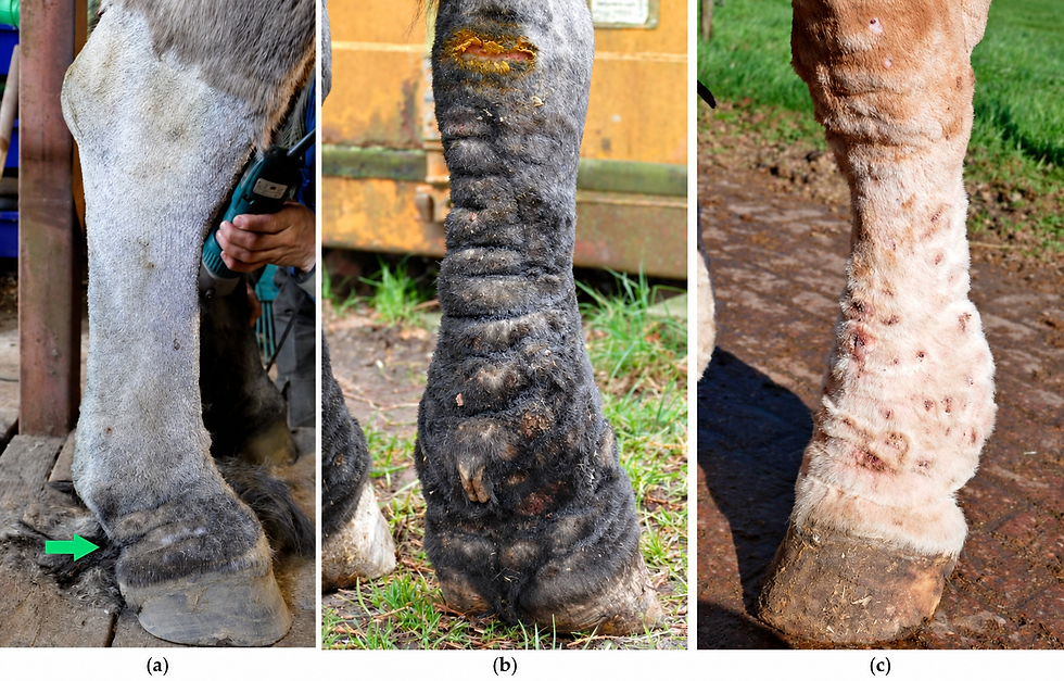
Mild or early CPL (a)
The prognosis for comfort may be fair to good with early recognition and consistent care. Swelling, itching, crusting, and infection may improve, but susceptibility remains.
Moderate CPL (b)
The long-term prognosis is guarded. Horses may remain useful and comfortable, but flare-ups are common and fibrosis generally continues to progress.
Advanced CPL (c)
The prognosis becomes guarded to poor with extensive fibrosis, large nodules, chronic ulcers, recurrent cellulitis, persistent pain, limited flexibility, lameness, or inability to tolerate hoof care. Humane euthanasia may become necessary when pain, infection, or loss of mobility cannot be controlled.
CPL is best understood as a manageable but presently incurable disease. Early intervention does not guarantee that progression will stop, but it gives the horse the best chance of retaining healthier skin, comfortable movement, and a useful quality of life.
CPL and Breeding Decisions
The inherited component of CPL should be considered before breeding. A young horse without visible lesions is not automatically free of risk because signs become clearer with age and may be hidden by feather.
Breeding decisions in small populations must also protect genetic diversity. Consider age, repeated examinations, family history, severity, pedigree information, and population health with guidance from breed organizations, veterinarians, or geneticists.
There is no definitive DNA test that can replace examination and informed selection. A horse should not be advertised as genetically clear of CPL based only on a normal-looking set of legs or an unrelated commercial genetic panel.
Conclusion
Chronic progressive lymphedema is a serious, lifelong condition of susceptible draft and heavily feathered horses. Impaired lymphatic clearance, inflammation, abnormal connective tissue, and fibrosis reinforce one another, while mites, infection, moisture, trauma, and reduced movement add further damage.
The most useful opportunity lies in early recognition. Part the feathers, palpate every surface of every lower limb, investigate recurring pastern problems, and keep objective records. Once disease is identified, careful skin access, complete drying, veterinary mite control, targeted treatment of infection, comfortable movement, wound protection, and regular hoof care can slow secondary damage and protect quality of life.
CPL management requires patience because the goal is control rather than cure. Small, consistent decisions made every day usually matter more than occasional intensive treatment.
Frequently Asked Questions About Chronic Progressive Lymphedema
1. What are the first signs of CPL in horses?
Early signs include mild persistent swelling, thickened or less mobile skin, a small fold behind the pastern, scaling beneath the feathers, recurrent crusts, and mild itching or stamping. The changes may be easier to feel than see. Part the hair and palpate the entire lower limb rather than judging it from a distance.
2. Is chronic progressive lymphedema the same as scratches or mud fever?
No. Scratches and mud fever are broad terms for pastern dermatitis and may result from moisture, friction, infection, mites, photosensitization, and other causes. CPL is a progressive disorder involving the lymphatic system, skin, and connective tissue. A horse with CPL may also develop pastern dermatitis as a secondary complication.
3. Can CPL in horses be cured?
There is currently no cure. Established fibrosis and severe lymphatic damage cannot be reliably reversed. Treatment focuses on slowing progression, reducing edema, controlling mites and infection, preserving skin integrity, maintaining movement, and protecting the horse's comfort.
4. Is CPL painful?
Early CPL may cause little obvious pain. As folds deepen and the skin becomes inflamed, infected, cracked, or ulcerated, the condition can become painful. Advanced fibrosis can also restrict movement and make lifting the feet difficult. Stamping or resistance to leg handling may reflect mites, pain, or both.
5. Should the feathers be clipped on a horse with CPL?
Clipping or shortening the feathers is often necessary to examine, clean, dry, and medicate active disease. It must be done carefully because the skin may be folded and fragile. Complete clipping is not required in every stable case, but appearance should never prevent access when mites, infection, crusting, or wounds are present.
6. Are feather mites the cause of CPL?
Chorioptes mites are strongly associated with CPL in some draft-horse populations and can greatly worsen itching, inflammation, wounds, and scaling. Current evidence does not establish them as the only cause. CPL has an inherited and tissue-level basis, while mites act as an important contributor and disease amplifier.
7. Can I treat Chorioptes mites with cattle pour-on medication?
Do not apply a livestock pour-on or calculate a treatment from an online study without veterinary direction. Some products are used off label, dose and route matter, and certain livestock acaricides are unsafe for horses. Amitraz should not be used on horses because of the risk of serious toxicity.
8. Does exercise help a horse with CPL?
Comfortable, regular movement can support lymphatic return and help preserve flexibility. It does not remove established fibrosis. Exercise must be adapted when the horse has lameness, acute cellulitis, deep infection, open wounds, arthritis, or severe pain. Ask the veterinarian what level of turnout or controlled work is appropriate.
9. Can a horse with CPL still be ridden or driven?
Some horses with mild or well-managed CPL remain capable of riding, driving, or other work. Suitability depends on soundness, skin integrity, limb flexibility, fitness, and comfort. Work should be reduced or stopped if the horse becomes lame, painful, develops an acute infection, or cannot move freely.
10. Should a horse with CPL be bred?
CPL has a substantial inherited component, so breeding an affected horse may pass increased susceptibility into the next generation. There is no definitive DNA test, and young horses can appear normal before signs develop. Decisions should consider clinical severity, age, family history, breed diversity, and veterinary or genetic advice.
Related Horse Education Online Resources
Selected Peer-Reviewed References
Brys M, Claerebout E, Chiers K. Chronic progressive lymphedema in Belgian Draft horses: understanding and managing a challenging disease. Veterinary Sciences. 2023;10:347. https://doi.org/10.3390/vetsci10050347
Brys M, Claerebout E, Chiers K. Alleviating lesions of chronic progressive lymphedema in Belgian Draft horses by successfully treating Chorioptes bovis infestation with moxidectin 0.5% pour-on. Veterinary Parasitology. 2023;324:110074. https://doi.org/10.1016/j.vetpar.2023.110074
Brys M, Claerebout E, Saey V, Chiers K. High prevalence of Chorioptes bovis: an important factor in chronic progressive lymphedema in Belgian Draft horses. Veterinary Research Communications. 2025;49:129. https://doi.org/10.1007/s11259-025-10695-y
De Cock HEV, Affolter VK, Wisner ER, Larson RF, Ferraro GL. Lymphoscintigraphy of draught horses with chronic progressive lymphoedema. Equine Veterinary Journal. 2006;38:148-151. https://doi.org/10.2746/042516406776563297
De Cock HEV, Affolter VK, Farver TB, Van Brantegem L, Scheuch B, Ferraro GL. Measurement of skin desmosine as an indicator of altered cutaneous elastin in draft horses with chronic progressive lymphedema. Lymphatic Research and Biology. 2006;4:67-72. https://doi.org/10.1089/lrb.2006.4.67
Mittmann EH, Mömke S, Distl O. Whole-genome scan identifies quantitative trait loci for chronic pastern dermatitis in German draft horses. Mammalian Genome. 2010;21:95-103. https://doi.org/10.1007/s00335-009-9244-z
Powell H, Affolter VK. Combined decongestive therapy including equine manual lymph drainage to assist management of chronic progressive lymphoedema in draught horses. Equine Veterinary Education. 2012;24:81-89. https://doi.org/10.1111/j.2042-3292.2011.00311.x
Sievers J, Distl O. The estimation of genetic parameters for chronic progressive lymphedema and body traits in the Rhenish German Draught Horse. Animals. 2024;14:1214. https://doi.org/10.3390/ani14081214


Comments